Journal of Signal and Information Processing
Vol. 4 No. 1 (2013) , Article ID: 28305 , 6 pages DOI:10.4236/jsip.2013.41010
Adaptive Gradient-Based and Anisotropic Diffusion Equation Filtering Algorithm for Microscopic Image Preprocessing
![]()
Jingling Institute of Technology, College of Information Technology, Nanjing, China.
Email: hailingliu1979@163.com
Received November 26th, 2012; revised December 28th, 2012; accepted January 9th, 2013
Keywords: Microscopic Image; Image Preprocessing; Anisotropic; Gradient-Based
ABSTRACT
In image acquisition process, the quality of microscopic images will be degraded by electrical noise, quantizing noise, light illumination etc. Hence, image preprocessing is necessary and important to improve the quality. The background noise and pulse noise are two common types of noise existing in microscopic images. In this paper, a gradient-based anisotropic filtering algorithm was proposed, which can filter out the background noise while preserve object boundary effectively. The filtering performance was evaluated by comparing that with some other filtering algorithms.
1. Introduction
1.1. Quality of Microscopic Images
The limitation of the quality of microscopic images can be attributed to image intensity levels, image resolution, image pixel size and dynamic ranges. The former two factors are related to the optical system and the latter two are related to the detector system.
1.2. Noise in Microscopic Images
Due to the influence of image quantization, adding quantization noises exist in microscopic images and the whole image background shows nonuniform distribution. In Figure 1, the gray level distributions of three horizontal areas were computed in three different positions Y = 30, 1215 and 1900. It is evident that the gray level distribution in image is not constant and nonuniform exists, which is smaller in background regions and larger in boundary regions. Another characteristic of microscopic image is that objects are always in motion. Due to the amplification effect of microscope, the image is sensitive to focal length and usually the images appear blurry near the boundaries and halo-type boundaries appear. In this case, the precise detection of boundary becomes very difficult. Figure 2 demonstrates three typical morphologies of cells in the image, which all have blurry and halotype boundaries. Because microscopic images are fluid images, apart from the objects, there exists some other objects like bubbles, sediments etc.
1.3. Microscopic Image Preprocessing Research Status
Given the complexity of factors introducing noise and intensity heterogeneity, there are two approaches for improving the quality of microscopic images. First, one could focus on improving specimen preparation and imaging conditions, for example improved fluorescent dyes. Second, one could attempt to correct pixel intensities after image acquisition, as it is the case for image restoration. Apart from some traditional filtering algorithm such as self-adaptive median filtering algorithm [1,2], hybrid filtering algorithm [3], anisotropic filtering algorithm [4], feature fusion filtering algorithm [5-7], wavelet based filtering algorithm [8-11] etc., some specific filtering and restoration algorithms were also proposed such as the empirical correction methods for intensity loss [12], constant thresholding [13], iterative correction methods [14], 2D histogram or estimations of intensity decay function [15]. Although these methods work well for certain category of microscopic image, most of them require the image meet some basic requirements such as the photo bleaching is a spatially homogeneous in a lateral plane, which greatly limit the universality and availability.
1.4. Quality Degration Model of Microscopic Images
Normally in image denoising techniques, noise can be
 (a)
(a) (b)
(b) (c)
(c)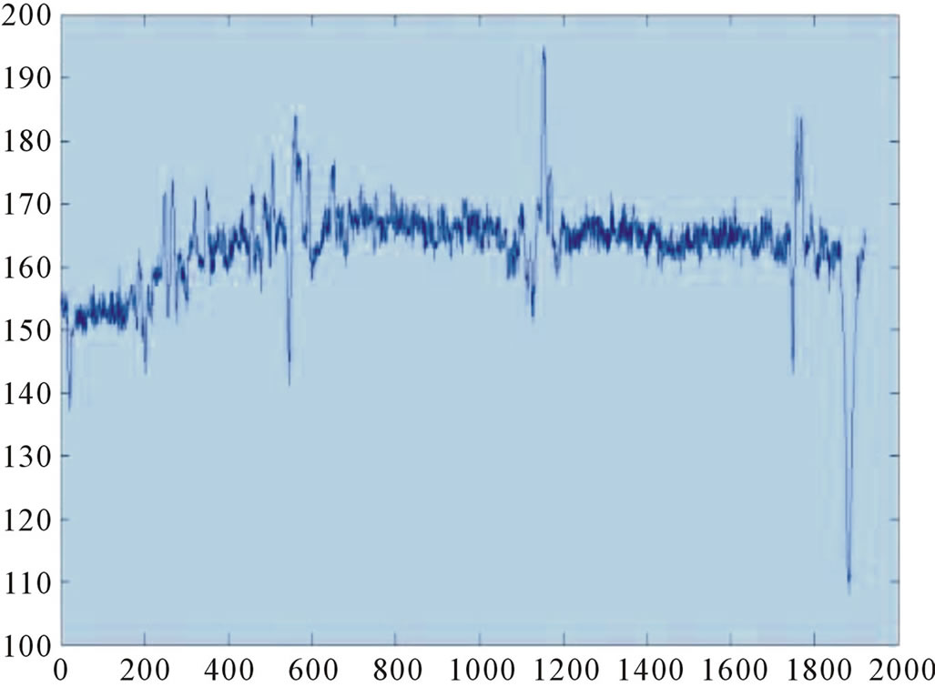 (d)
(d)
Figure 1. Graylevel distribution at different parts of microscopic image. (a) Scanning lines; (b) Y = 30; (c) Y = 1215; (d) Y = 1900.
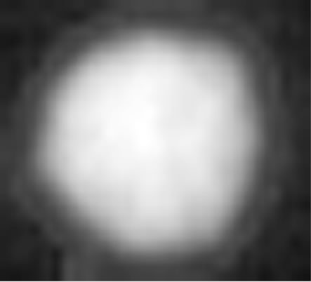
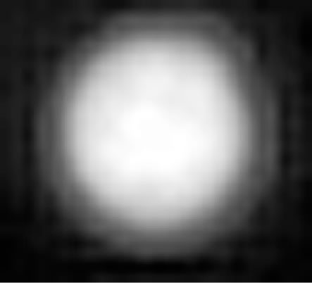

Figure 2. Cells with blurry boundaries.
ideally divided into adding noise and multiplicative noise. Suppose the degraded image as g, the ideal image as f, and the noise as n. The adding model and the multiplicative model can be described in formula (1) and (2). Background noise in microscopic images can normally be regarded as adding Gaussian noise and low magnification lens tiny particles as adding pulse noise.
 (1)
(1)
 (2)
(2)
Besides noises, there are some other quality degradation factors like inappropriate focusing, uneven illumination etc. For better cell segmentation and recognition, the system should design a good preprocessing algorithm to overcome noise and other image quality degration factors.
The boundary of cell is very important for quantitative analysis of microscopic images. As described above, the qualities of microscopic image are always degraded by adding noise and halo-type boundaries. So it is very important to design an appropriate filtering algorithm that can filter out the noise and will not further degrade the image quality of boundaries. In this paper, an adaptive gradient-base and anisotropic diffusion equation filtering algorithm was proposed to deal with this problem.
2. Proposed Algorithm
In order to accelerate the filtering speed, a judgment should be made to each pixel in image and then the filtering manipulation is implemented on pixels that are judged as noises. For non-noise pixel, no filtering manipulation is implemented.
2.1. Gradient Based Background Noise Detection
Based on the description in introduction section, it is known that in background area, the gray scale changes less while in boundary area or noise it changes more. Therefore, we can compute the variation of gray scale in a sliding window  while it slides through the whole image to judge the property of each pixel, boundary or noise. Then the filtering is implemented on possible noise candidate pixel. Suppose the gradient of each direction as
 , i denotes the all eight directions. The judgment rule is described as following equations:
 (3)
(3)
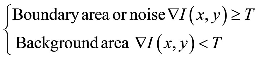 (4)
(4)
After the judgment is made for all pixels, the filtering manipulation is implemented on the pixels that are judged as boundary or noise.
2.2. Anisotropic Filtering Algorithm
According to the concept of divergence field, anisotropic diffusion equation can be described in the following equation:
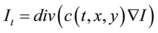 (5)
(5)
In Equation (5),  is divergence operator,
  is gradient operator, I is the function of
 ,
, is spatial scale function, namely the diffusion coefficient, which is a nonnegative monotonic decreasing function, Ii is the derivative of I with respect to time t, t is the time of thermal diffusion.
The selection of diffusion coefficient  will directly influence the filtering effect and in general, diffusion coefficient
  is given a value of the norm of a vector, which has the following attributes:
1) In interior area .
.
2) In the boundary area,  ,
,  is the unit vector of the gradient of point
  in the boundary area.
  is the strength difference at each side of the boundary.
We can determine  as
as
 (6)
(6)
The classical  can be described as
 (7)
(7)
Or
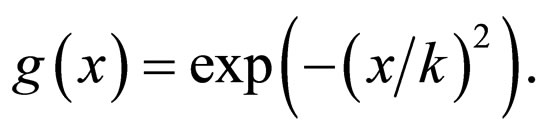 (8)
(8)
Here, k is the controlment coefficient of diffusion strength, x is the gradient of the diffusion point. According to the definition of diffusion coefficient, in different directions different coefficients are adopted. In addition, the monotonic decreasing function was taken in different directions as the diffusion coefficient. In background area or interior area of image, the gray level values are similar and gradient is very small, so the diffusion coefficient is large to implement smoothing. On the contrary, in the area of boundary, gray level value changes much and accordingly the gradient increases, diffusion coefficient is very small to preserve the boundary information. Here we consider the processing in eight directions. Figure 3 demonstrate the flow chart of the proposed algorithm.
The algorithm is achieved using iterations that are described in Table 1 as the discretization of the thermal diffusion process. The detailed algorithms are as follows.
3. Experimental Results
The filtering was implemented using the proposed algorithm of gradient-based background noise detection and anisotropic filtering. The diffusion coefficient is set as 10, iteration step size set as 0.21, iteration time as 5, 10, 15, 20 and thresholding value for boundary and noise as 30.
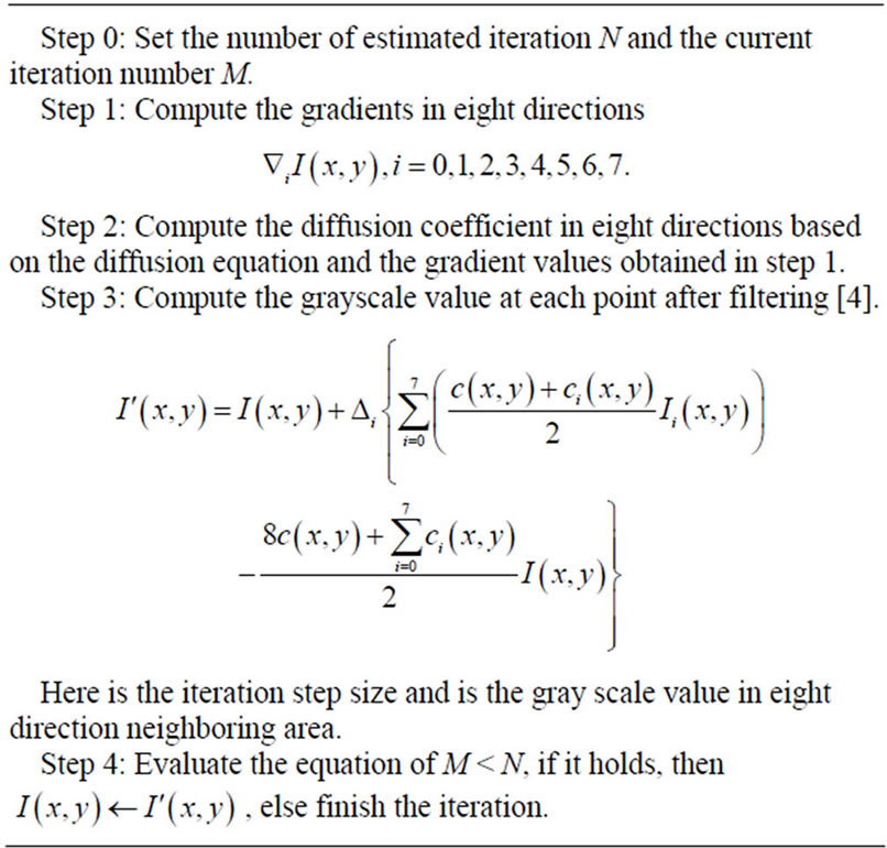
Table 1. The discretization process of the proposed algorithm.
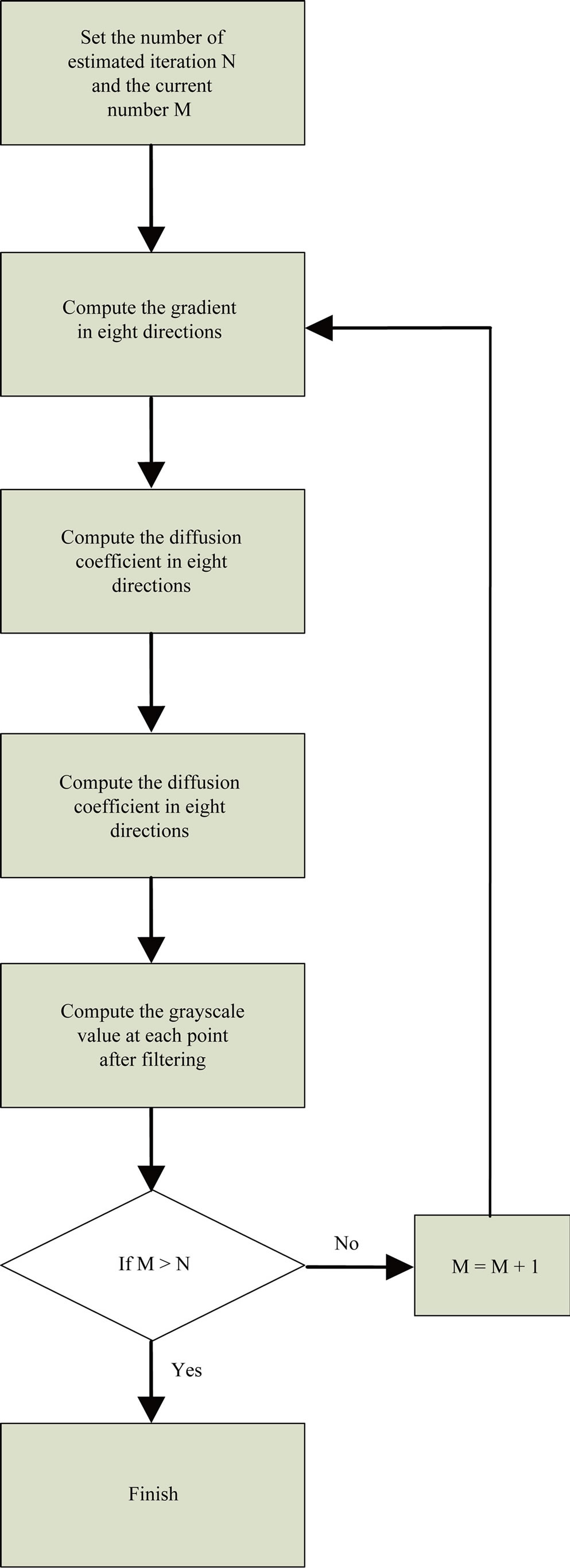
Figure 3. Flow chart of proposed algorithm.
A sample microscopic image was used, which incorporates the typical features of microscopic images with adding background noise, pulse noise and tiny particle noise. The results of the algorithm are compared with that of median filtering. It is very clear that the filtering algorithm proposed above can obtain satisfactory results in removing background noise as well as preserving object boundaries. In addition, by using noise detection algorithm, the computational speed improved by 3 times compared with using anisotropic filtering algorithm alone.
Figure 4 demonstrates the comparison of experimental results between the proposed algorithm and the median filter. The top row shows the filtering results of the proposed algorithm using different iteration times. The bottom row shows the filtering results of traditional median filtering algorithm using different padding sizes. It is evident that our algorithm obtained better performance in removing noises while preserving boundary details.
To demonstrate the advantages of the proposed algorithm to other filtering algorithm, the performance of proposed algorithm (Aniso) was compared with other four widely used filtering algorithms in medical image processing including homomorphic filtering algorithm (Homo) [16], DCT-based filtering algorithm [17], wavelet based filtering algorithm (Wavelet) [18], isotropic diffusion based filtering algorithm (Iso) [19]. The results in Figure 5 demonstrate that the proposed algorithm outperforms other algorithms in filtering out noise while preserving boundaries.
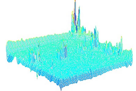
Figure 6 demonstrates the surface map of the original image and the filtered image using proposed algorithm and other four algorithms. Surface map draws a wire frame mesh with color determined by the intensity value in the image and the color is proportional to the surface height. It is evident that the proposed anisotropic diffusion filtering algorithm can obtain better filtering performance in removing noise and preserving boundary
 (a)
(a) (b)
(b)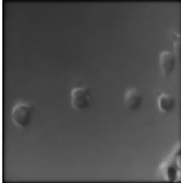 (c)
(c)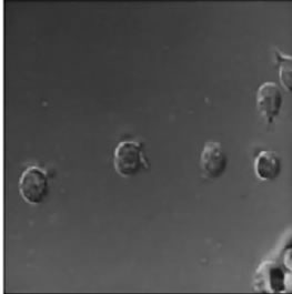 (d)
(d)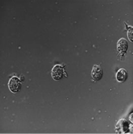 (e)
(e)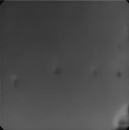 (f)
(f) (g)
(g)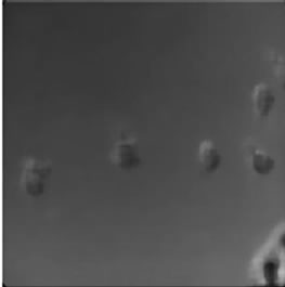 (h)
(h)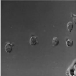 (i)
(i)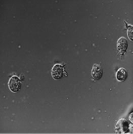 (j)
(j)
Figure 4. Filtering performance comparison between proposed algorithm and median filter. (a) Original; (b) T = 5; (c) T = 10; (d) T = 20; (e) T = 30; (f) Original; (g) S = 4; (h) S = 6; (i) S = 10; (j) S = 12.
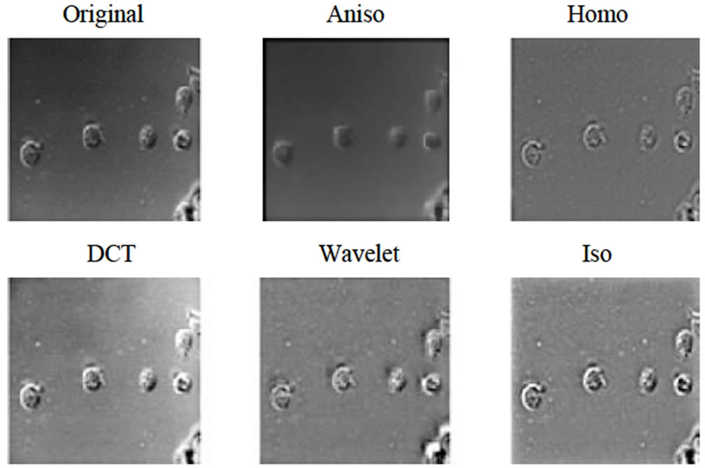
Figure 5. Comparison of different filtering algorithm.
 (a)
(a)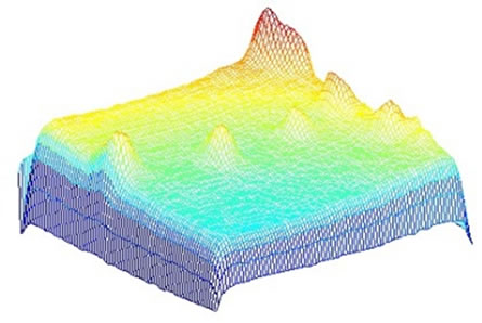 (b)
(b)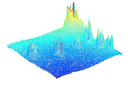 (c)
(c)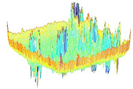 (d)
(d) (e)
(e)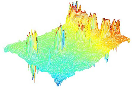 (f)
(f)
Figure 6. Comparison of surface map of different filtering algorithm. (a) Original; (b) Aniso; (c) Homo; (d) DCT; (e) Wavelet; (f) Iso.
information while the performance of other four algorithms are not very satisfactory in our experiment.
4. Conclusion
In this paper, an adaptive gradient-based and anisotropic diffusion equation filtering was proposed to filter the noise of cell image. The algorithm was proposed to preprocess microscopic images, which can be divided into two steps: 1) the judgment of pixels, background area or boundary area; 2) anisotropic filtering using different coefficient. The experimental results demonstrate that the proposed algorithm outperforms other filtering algorithms in microscopic image preprocessing, which can filter out noises while preserving the boundaries effectively.
REFERENCES
- V. Khryashchev and A. L. Priorov, “Image Denoising Using Adaptive Switching Median Filter,” IEEE International Conference on Image Processing, Vol. 1, 2005, pp. 117-120.
- K. NallaperUmal, J. Varghese and S. Saudia, “Adaptive Rank-Ordered Switching Median Filter for Salt & Pepper Impulse Noise Reduction,” IEEE Annual India Conference, New Delhi, 15-17 September 2006, pp. 1-6.
- Y. C. Wang, D. Q. Liang, H. Ma and Y. Wang, “An Algorithm for Image Denoising Based on Mixed Filter,” Proceedings of the 6th World Congress on Intelligent Control and Automation, Vol. 2, 2006, pp. 9690-9693.
- Y. J. Yu and S. T. Acton, “Speckle Reducing Anisotropic Diffusion,” IEEE Transaction on Image Processing, Vol. 11, No. 11, 2002, pp. 1260-1269. doi:10.1109/TIP.2002.804276
- M. E. Izquierdo and M. Ghanbari, “Texture Smoothing and Object Segmentation Using Feature-Adaptive Weighted Gaussian Filtering,” SBT/IEEE International Telecommunications Symposium, Sao Paulo, 9-13 August 1998, pp. 650-655.
- M. Basu, “Gaussian-Based Edge-Detection Methods—A Survey,” IEEE Transactions on System, Man and Cybernetics, Vol. 3, No. 32, 2002, pp. 252-260.
- S. J. Fu, Q. Q. Ruan, Y. L. Geng and W. Q. Wang, “Feature-Oriented Coupled Bidirection Flow for Image Denoising and Edge Sharping,” Tencon 2005 IEEE Region, Melbourne, 21-24 November 2005, pp. 1-5.
- Y. Zhen, “Wavelet Domain Multiresolution Markov Models for Image Segmentation and Denoising Applications,” Proquest Information and Learning Company, 2005, pp. 78-90.
- J. Portilla and V. Strela, “Image Denoising Using Scaled Mixtures of Gaussians in the Wavelet Domain,” IEEE Transactions on Image Processing, Vol. 11, No. 12, 2004, pp. 1338-1351.
- J. Liu and P. Moulin, “Image Denoising Based on Scale-Space Mixture Modeling of Wavelet Coefficient,” IEEE International Conference on Image Processing, Vol. 1, 1999, pp. 386-390.
- C. Dongwook and T. D. Bui, “Multivariate Statistical Approach for Image Denoising,” IEEE International Conference on Acoustics, Speech and Signal Processing, Vol. 4, 2005, pp. 589-592.
- K. Rodenacker and P. Aubele, “Groping for Quantitative Digital 3-D Image Analysis: An Approach to Quantitative Fluorescence in Situ Hybridization in Thick Tissue Sections of Prostate Carcinoma,” Analytical Cellular Pathology, Vol. 15, No. 1, 1997, pp. 19-29.
- T. Irinopoulo, J. Vassy, M. Beil, P. Nicolopoulo and D. Encaoua, “3-D DNA Image Cytometry by Confocal Scanning Lasermicroscopy in Thick Tissue Blocks of Prostatic Lesions,” Cytometry, Vol. 27, No. 2, 1997, pp. 99-105. doi:10.1002/(SICI)1097-0320(19970201)27:2<99::AID-CYTO1>3.0.CO;2-F
- T. Visser, F. Groen and G. Brakenhoff, “Absorption and Scattering Correction in Fluorescence Confocal Microscopy,” Journal of Microscopy, Vol. 163, No. 2, 2007, pp. 189-200. doi:10.1111/j.1365-2818.1991.tb03171.x
- A. Liljeborg, M. Czader and A. Porwit, “A Method to Compensate for Light Attenuation with Depth in 3D DNA Image Cytometry Using a Confocal Scanning Laser Microscope,” Journal of Microscopy, Vol. 177, No. 2, 1995, pp. 108-114. doi:10.1111/j.1365-2818.1995.tb03540.x
- S. E. Umbaugh, “Computer Imaging: Digital Image Analysis and Processing,” CRC Press, Florida, 2005
- L. Yaroslavsky and M. Eden, “Fundamentals of Digital Optics,” Birkhauser, Boston, 1996. doi:10.1007/978-1-4612-0845-7
- T. Zhang, B. Fang, Y. Yuan, Y. Y. Tang, Z. Shang, D. Li and F. Lang, “Multiscale Facial Structure Representation for Face Under Varying Illumination,” Pattern Recognition, Vol. 42, No. 2, 2009, pp. 252-258.
- P. Perona, “Steerable-Scalable Kernels for Edge Detection and Junction Analysis,” Image and Vision Computing, Vol. 10, No. 10, 1992, pp. 663-672. doi:10.1016/0262-8856(92)90011-Q