Journal of Biophysical Chemistry
Vol.07 No.04(2016), Article ID:70670,11 pages
10.4236/jbpc.2016.74008
Mitochondrial Respiration Is Associated with Alloxan-Induced Mitochondrial Permeability Transition
Koichi Sakurai*, Mika Itoh
Department of Life Science, Hokkaido Pharmaceutical University School of Pharmacy, Sapporo, Japan

Copyright © 2016 by authors and Scientific Research Publishing Inc.
This work is licensed under the Creative Commons Attribution International License (CC BY 4.0).
http://creativecommons.org/licenses/by/4.0/


Received: August 15, 2016; Accepted: September 14, 2016; Published: September 19, 2016
ABSTRACT
We previously showed that increased mitochondrial inner membrane permeability which is known as mitochondrial permeability transition (MPT) is triggered by adding succinate in the presence of the diabetogenic agent alloxan. Here, our aim was to investigate whether mitochondrial respiration is associated with alloxan-induced MPT. After mitochondria isolated from rat liver were incubated with alloxan at 37˚C for 5 min, the addition of succinate immediately triggered the MPT in the presence of rotenone. However, little or no induction occurred at incubation temperatures below 25˚C. Malate/glutamate also triggered MPT by alloxan in the absence of rotenone. In mitochondrial suspensions containing alloxan, succinate accelerated oxygen consumption that was completely inhibited by cyanide. These results suggest that mitochondrial respiration is associated with the alloxan-induced MPT. Alloxan radical production was investigated using ESR spectroscopy. Mitochondria incubated with succinate and alloxan elicited low signal intensity (radical formation) that increased significantly in the presence of cyanide. When the incubation of alloxan with mitochondria after the addition of succinate, a little intensity of the signal was observed, but it was remarkably increased after the addition of cyanide. Ubiquinone analogues inhibited the MPT induction. These results suggest that the initiation of MPT is associated with alloxan redox cycling via an electron transfer process at a quinone-binding site in respiratory mitochondria.
Keywords:
Mitochondria, Permeability, Respiration, Alloxan, Redox Cycle, Radical

1. Introduction
Mitochondria are known to regulate apoptotic cell death, in addition to their critical function in energy metabolism. Loss of mitochondrial integrity is a causative event, leading to apoptosis in cells after oxidative stress, Ca2+ toxicity and ischemia/reperfu- sion, thus supporting the idea that mitochondrial permeability transition (MPT) lead to the initiation of programmed cell death, apoptosis [1] - [3] . This MPT is thought to implicate in cellular Ca2+ homeostasis, import of protein into mitochondria, and thermal regulation in mammalian mitochondria. An assortment of agents can induce the MPT due to the opening of a large pore that allows components with a molecular mass of 1.5 kDa or lower to diffuse across the inner membrane [4] - [7] . The quinoidal compound alloxan has diabetogenic action. In previous reports we demonstrated that alloxan induced apoptotic death in INS-1 cells (a rat pancreatic β-cell line) and that the coincident mitochondrial damage is linked to apoptosis [8] , and that alloxan induces MPT that requires mitochondrial energization, oxidation of protein thiols, and matrix ATP to promote energized uptake of Ca2+ [9] . Several studies have demonstrated theinvolve- ment of mitochondrial events such as activation of mitochondrial-derived factors and the decrease in mitochondrial transmembrane potential in apoptosis [10] [11] . Ubiquinone is a respiratory chain component buried within the inner membrane where it accepts two electrons from complexes I or II. The reduced adduct, termed ubiquinol, then donates electrons to complex III. While ubiquinone and its analogs can regulate MPT [12] [13] , the role of mitochondrial respiration in the induction of MPT is not clear.
Several researchers have demonstrated that the diabetogenic action of alloxan is initiated by the generation of reactive oxygen species (ROS) [9] [14] - [16] , althoughthe detailed molecular mechanism of alloxan-mediated cytotoxicity is not yet clearly understood. The initial step for ROS generation is mostly mediated by the mitochondrial electron transfer system [17] . Alloxan is a mild oxidant that is easily reduced to alloxan radical by GSH or ascorbate [18] [19] . MPT can be induced by a diverse range of stimuli, including prooxidants, ganglioside GD3, fatty acids, quinone compounds and thiol oxidants [10] [12] [20] [21] . Here, we showed that mitochondrial respiration is associated with alloxan-induced MPT triggered by succinate, and that MPT should be associated with redox cycling of alloxan via an electron transfer process occurring at a quinone binding site in respiratory mitochondria.
2. Materials and Methods
2.1. Materials
Alloxan and sodium succinate were purchased from Wako Pure Chemical Industries Ltd., Osaka, Japan. SOD (from bovine erythrocytes) and catalase (from bovine liver, thymol free) were from Sigma Co., St. Louis, MO. Other chemicals were of analytical grade from commercial supplies.
2.2. Measurement of MPT
Liver mitochondria were prepared daily from male Wistar-strain rats weighing ~200 g that were fasted overnight as described previously [22] . All rats were treated in accordance with the guidelines of Hokkaido College of pharmacy for the treatment of animals. To determine the integrity of mitochondria, State 3 respiration and State 4 respiration were measured by the method of Chance and Williams [23] . The averaged respiratory control ratio (State 3/State 4) of control mitochondria was 5.5 ± 0.2 for succinate as the substrate, suggesting the integrity of mitochondria. The standard experimental conditions for MPT were as follows: mitochondria (1 mg/ml of protein) were equilibrated at 37˚C for 5 min in a total volume of 3 ml of Chelex 100 resin-treated medium containing 10 mM Tris-HCl pH 7.4, 0.25 M sucrose, and 0.1 μM rotenone. The MPT was triggered by adding 5 mM succinate to this suspension. The permeability transition of mitochondria resulting from permeation of sucrose into the mitochondrial matrix was measured by recording the decrease in absorbance at 540 nm. Various inhibitors were included at the beginning of the preincubation period before adding alloxan.
2.3. Measurement of Oxygen Consumption
Oxygen consumption was measured by polarography at 37˚C using a Clark-type oxygen electrode (YSI model 5300, Yellow Springs Instrument Co., Inc., Yellow Springs, OH). Mitochondria (1 mg/ml of protein) were suspended in the buffer above containing 0.1 μM rotenone at 37˚C in the presence or absence of 1.0 mM alloxan. The reaction was triggered by the addition of 5.0 mM succinate.
2.4. Measurement of Radical Species
ESR spectra were obtained using a JEOL model JES RE1X (Tokyo, Japan). The standard incubation mixtures were the same as those for MPT studies. Spectrometer settings for the alloxan radical were as follows: Magnetic field, 3365G; microwave power, 5.0 mW; modulation frequency, 9.450 GHz; field modulation width, 0.20 G; time constant, 0.3 s and 2.5 G/min scan speed. After the incubation at 37˚C for the indicated times, samples were rapidly aspirated into the aqueous flat cell and the ESR spectra were recorded.
2.5. Statistical Analysis
Data are expressed as means ± S.E. and were statistically analyzed using the Student’s t test for paired data. p < 0.05 was considered statistically significant.
3. Results
3.1. Electron Transfer System Involved in MPT
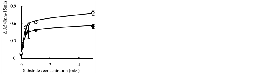
We previously reported that alloxan promotes enhanced ATP production in mitochondria in the presence of succinate [9] . We therefore carried out additional studies to evaluate whether mitochondrial respiratory function is associated with alloxan-induced MPT in isolated rat liver mitochondria. Mitochondria were preincubated with alloxan at 37˚C for 5 min in the presence or absence of rotenone, and MPT was then triggered by adding succinate or malate/glutamate, respectively (Figure 1(a)). Addition of either malate/glutamate (<0.3 mM/0.3 mM) in the absence of rotenone or succinate (<0.5 mM) in the presence of rotenone clearly promoted MPT in a dose-dependent manner

 (a) (b)
Figure 1. Alloxan-induced MPT triggered by the addition of respiratory substrates. (a) After mitochondria were preincubated with 1 mM alloxan for 5 min at 37˚C, MPT was triggered by adding respiratory substrates: 1, alloxan; 2, 0.5 mM malate/0.5 mM glutamate or 5 mM succinate; 3, 1 + 0.5 mM malate/0.5 mM glutamate; and 4, 1 + 5 mM succinate. Closed and open arrows indicate the addition of alloxan and respiratory substrates, respectively. Typical results of one experiment are shown, and similar results were obtained with at least three different preparations of mitochondria; (b) after preincubation with 1 mM alloxan for 5 min, MPT was triggered by adding various concentrations of malate/glutamate (closed circles) or succinate (open circles). Data are expressed as an average of triplicate experiments ± S.E. (error bars)
(Figure 1(b)). Figure 2 shows the effect of incubation temperature on alloxan-induced MPT triggered by succinate. MPT was clearly observed at incubation temperatures above 25˚C with the maximum rate being achieved at ~37˚C. However, a decline of MPT was observed at 45˚C. MPT was not observed with either respiratory substrate in mitochondria preincubated without alloxan. These results indicate that respiratory substrates are necessary for the initiation of alloxan-induced MPT.
To confirm the involvement of mitochondrial respiration in the induction of MPT, oxygen consumption was measured in mitochondria treated with alloxan and/or succinate which is substrate involved in donating electrons to complex II. The rate of mitochondrial oxygen consumption increased slightly upon addition of alloxan (Figure 3). Remarkably, the addition of succinate also elicited increased oxygen consumption. KCN almost completely inhibited the increase in oxygen consumption, so did NaN3. In the presence of rotenone, the oxygen consumption by addition of succinate as respiratory substrate was strongly inhibited by the complex II inhibitor, thenoyltrifluoroacetone, and the complex III inhibitor, antimycin A (data not shown). These results suggest that the oxygen consumption resulted from mitochondrial respiration. Addition of succinate to mitochondria prior to alloxan caused rapid onset of oxygen consumption, and subsequent addition of alloxan resulted in a slight decrease in the consumption rate. Interestingly, KCN scarcely inhibited consumption under these circumstances. These results suggest that mitochondrial respiration is associated with MPT induction in mitochondria preincubated with alloxan.
3.2. Genertiopn of Alloxan Radical
Alloxan has a quinone-like structure that is easily converted to the alloxan radical by

Figure 2. Effect of incubation temperature on alloxan-induced MPT. Before adding alloxan, mitochondria (1 mg/ml of protein) were equilibrated for 5 min in a total volume of 3 ml of medium consisting of 10 mM of Tris-HCl, 0.25 M sucrose, and 0.1 μM rotenone at the indicated temperatures. Other conditions were as described in Materials and Methods. Each value is the mean ± S.E. of triplicate experiments.
 (a) (b)
Figure 3. Effect of alloxan on the respiration in mitochondria incubated with succinate. Alloxan (1 mM) was added to the mitochondrial suspension (1 mg/ml of protein) before (a) or after (b) adding 5 mM succinate. Each value in parentheses represents the mean ± S.E. of triplicate experiments. Typical results of one experiment are shown, and similar results were obtained in three other independent experiments using three different preparations of mitochondria.
univalent reduction [18] [19] . To investigate whether the redox state of alloxan induced by the respiration has been implicated in the initiation of the MPT, the generation of alloxan radical in mitochondria was determined using ESR spectroscopy (Figure 4). After the addition of succinate, no ESR spectrum could be clearly detected in mitochondria preincubated with alloxan (spectrum 1). The addition of KCN resulted in an

Figure 4. Generation of the alloxan radical in mitochondria. The reaction mixture consisted of mitochondria (1 mg/ml of protein), 1 mM alloxan and 5 mM succinate with or without 0.3 mM KCN. The generation of the alloxan radical was detected as described in Materials and Methods. Spectrum 1, The ESR signal was determined at 1 min after the addition of succinate on mitochondria pretreated with alloxan. 2, KCN was added at 1 min after adding succinate under the conditions in spectrum 1, and then the spectrum was determined immediately. 3, KCN was present from the beginning of the incubation. Other conditions were as described for spectrum 1. 4, Simulated ESR spectrum using the following parameters: hyperfine coupling constant of aH = aN = 0.45 G. The spectrum matched well with the experimental results. Typical results of one experiment are shown, and similar results were obtained in at least three other independent experiments.
increase in ESR signal intensity under the same conditions (spectrum 2). The ESR spectrum obtained here (in the presence of KCN) was identified as the alloxan radical from the hyperfine constants aN = aH = 0.045 mT and the g-value = 2.0052, in agreement with the simulated spectrum (spectrum 4) and our previous spectra [18] [19] . When KCN was present from the beginning of the incubation, the intensity of signal markedly decreased (spectrum 3). A low level of alloxan radical was observed by the addition of antimycin A in respiring mitochondria (data not shown). These results suggest that the alloxan redox cycle is supported in respiring mitochondria.
3.3. Effect of Quinone Cycle Antagonists and Ubiquinol Analogs on MPT
Given that alloxan has a quinone-like structure, we next investigated the interaction of alloxan with quinone-binding sites during the induction of MPT. Antagonists (myxothiazol and stigmatellin) of a quinol oxidation/proton output (Qo) site of the bc1 complex in the quinone (Q) cycle almost completely inhibited alloxan-induced MPT; and significant inhibition was also evident with the antagonist antimycin A acting at a quinone reduction/proton input (Qi) site of the bc1 complex (Table 1). It has been proposed that quinone analogs modulate the Ca2+-dependent MPT pore through a common binding site [12] [13] . Ubiquinone 0 and decylubiquinone significantly inhibited alloxan-induced MPT (Table 1). In the absence of alloxan, none of the inhibitors and analogs used in this study had an effect on the permeability of mitochondria and the absorbance at 540 nm in the same range of concentrations effective in inhibiting alloxan-induced MPT (data not shown). These results suggest that alloxan might induce MPT via interaction with the quinone-binding site.
3.4. Effect of Antioxidants on MPT
Because the alloxan radical reduces molecular oxygen to the superoxide anion radical (O2•−), we investigated the involvement of ROS in the MPT induced by alloxan. As shown in Table 2, the antioxidant trolox, the hydroxyl radical scavengers thiourea and DMSO, and the antioxidant enzymes SOD or catalase did not inhibit the MPT. Furthermore, no decrease in intensity of the ESR signal was observed when any antioxidants were added to the alloxan radical-generation system of alloxan with GSH (data not shown). These results suggest the possibility that alloxan radical generated was directly involved in MPT induction, rather than ROS.

Table 1. Effects of antagonists of the quinone cycle and ubiquinone analogs on alloxan-induced MPT.
Experimental conditions were as described in Figure 2, except that antagonists of the Q cycle and ubiquinone analogs were present from the beginning of the incubation. Each value represents the mean ± S.E. of triplicate experiments.
Table 2. Effect of antioxidants on alloxan-induced MPT.
Experimental conditions were as described in Figure 2, except that antioxidants were present from the beginning of the incubation. Each value represents the mean ± S.E. of triplicate experiments.
4. Discussion
The rate and extent of MPT is dependent on mitochondrial ATP content [9] . The present work was carried out to test whether mitochondrial respiration participates in alloxan-induced MPT. We demonstrated that the rate and extent of MPT increases with increasing incubation temperature in that alloxan-induced MPT was initiated above 25˚C and reached the maximum rate at ~37˚C. In the presence of alloxan, the rate of mitochondrial respiration increased significantly after addition of succinate (compared to that in the absence of alloxan), which was almost completely inhibited by KCN. Furthermore, the addition of malate/glutamate as well as succinate triggered alloxan-in- duced MPT, and that was inhibited by the inhibitors of complex III center o, myxothiazol and stigmatellin, and inhibitor of complex III center I, antimycin A. We demonstrated the complex IV inhibitor KCN inhibited alloxan-induced MPT triggered by succinate [9] . These results suggest that the interaction between alloxan and the mitochondrial electron transfer system is involved in the induction of MPT.
When alloxan was added after succinate, only a low level of alloxan radical was observed. It's probably difficult that alloxan enters the intermembrane space in energized mitochondria, since alloxan has pK 6.63. Under the same condition, the subsequent addition of KCN did not increase the signal intensity of the alloxan radical. The addition of KCN to respiring mitochondria containing alloxan enabled us to detect the alloxan radical by ESR spectroscopy. The addition of KCN to mitochondria should elicit the reduced state of electron transfer system. Sugioka et al. demonstrated that the ubisemiquinone (a one-electron reduced form of coenzyme Q) could be generated in a succinate/fumarate coupled-KCN-submitochondrial system in the presence of molecular oxygen [24] . These results suggest that alloxan is reduced to the alloxan radical in energized mitochondria and that the alloxan redox cycle may participate in the induction of MPT. Despite the fact that respiration was almost completely inhibited by the addition of KCN, little effect of KCN on oxygen consumption was observed when alloxan was added after succinate. These results can be explained by assuming the reaction of alloxan and reduced substance such as sulfhydryl group in protein rather than the mitochondrial electron transport system [18] [19] .
The present study demonstrated that quinone analogs remarkably prevented MPT induced by alloxan which has a quinone-like structure. The ability of ubiquinone analogs to inhibit alloxan-induced MPT is probably associated with their capacity to bind a quinone-specific site. Fontaine et al. [12] demonstrated that quinones including ubiquinone analogs can elicit MPT, and suggested that a ubiquinone-binding site is directly involved in the regulation of the mitochondrial permeability transition pore. Ubiquinone is considered to act as a mobile electron carrier between complexes I, II and III in the mitochondrial electron transfer system, and Walter et al. [13] proposed that there are two specific sites with which quinone analogs could combine in this system. These exogenously added analogs can act as electron acceptor substrates for Complexes I and III [25] . Given that succinate-triggered MPT by alloxan is strongly inhibited by ubiquinone analogs as well as the fact that the rate and extent of MPT triggered by the addition of malate/glutamate or succinate/rotenone are similar, ubiquinone analogs may impart their inhibitory effect via binding to a quinone-specific site in Complex III, another site of ubiquinone binding site in complex I. From these findings, we speculated that alloxan bind to the affinity site for quinone in the electron transfer system.
It has been reported that MPT can be also induced by oxidative stress including thiol oxidation [9] [21] and the production of ROS in mitochondria may reduce the threshold of MPT by Ca2+ [26] . ROS can induce the MPT by targeting specific thiol residues of certain MPT components [27] . Mitochondria may be a significant source of ROS given that ~90% of the oxygen consumed by the cell is taken up by mitochondria and that dysfunction in the electron transfer system markedly enhances ROS production [28] [29] . In the present study, the MPT and oxygen consumption triggered by the addition of succinate in the presence of alloxan was strongly inhibited by the inhibitors of mitochondrial respiration such as KCN, NaN3, antimycin A and thenoyltrifluoroacetone. However, alloxan-induced MPT is not significantly prevented by exogenous SOD, catalase, or the classical •OH scavengers thiourea and DMSO. From these findings, we deduced that a leak of electron from mitochondrial electron transfer system caused the reduction of alloxan to alloxan radical, and that O2•−, H2O2 and •OH might not be responsible for the MPT.
5. Conclusion
In conclusion, the induction of MPT by alloxan appears to be dependent on mitochondrial respiration. It has been proposed that several mitochondrial disorders are associated with the onset of several diseases. Since the diabetogenic agent alloxan should interact with the quinone-binding site in Complex III, we presume that mitochondrial dysfunction may constitute one of the causal factors involved in pancreatic β-cell death. The results present here may be helpful in designing of new strategies to encourage further research efforts to aid in the development of therapeutic concept in diabetes mellitus.
Cite this paper
Sakurai, K. and Itoh, M. (2016) Mitochondrial Respira- tion Is Associated with Alloxan-Induced Mi- tochondrial Permeability Transition. Jour- nal of Biophysical Chemistry, 7, 87-97. http://dx.doi.org/10.4236/jbpc.2016.74008
References
- 1. Tsujimoto, Y. and Shimizu, S. (2007) Role of the Mitochondrial Membrane Permeability Transition in Cell Death. Apoptosis, 12, 835-840.
http://dx.doi.org/10.1007/s10495-006-0525-7 - 2. Green, D.R. and Reed, J.C. (1998) Mitochondria and Apoptosis. Science, 281, 1309-1312.
http://dx.doi.org/10.1126/science.281.5381.1309 - 3. Di Lisa, F., Carpi, A., Giorgio, V. and Bernardi, P. (2011) The Mitochondrial Permeability Transition Pore and Cyclophilin D in Cardioprotection. Biochimica et Biophysica Acta, 1813, 1316-1322.
http://dx.doi.org/10.1016/j.bbamcr.2011.01.031 - 4. Ichimura, T., Ito, M., Takahashi, K., Oyama, K. and Sakurai, K. (2011) Involvement of Mitochondrial Swelling in Cytochrome c Release from Mitochondria Treated with Calcium and Alloxan. Journal of Biophysical Chemistry, 2, 10-18.
http://dx.doi.org/10.4236/jbpc.2011.21002 - 5. Ruiz, L.M., Jensen, E.L., Bustos, R.I., Argüelloa, G., Gutierrez-Garcia, R., González, M., Hernández, C., Paredes, R., Simon, F., Riedel, C., Ferrick, D. and Elorza, A.A. (2014) Adaptive Responses of Mitochondria to Mild Copper Deprivation Involve Changes in Morphology, OXPHOS Remodeling and Bioenergetics. Journal of Cellular Physiology, 229, 607-619.
http://dx.doi.org/10.1002/jcp.24484 - 6. Bernardi, P. (1999) Mitochondrial Transport of Cations: Channels, Exchangers, and Permeability Transition. Physiological Reviews, 79, 1127-1155.
- 7. Penzo, D., Tagliapietra, C., Colonna, R., Petronilli, V. and Bernardi, P. (2002) Effects of Fatty Acids on Mitochondria: Implications for Cell Death. Biochimica et Biophysica Acta, 1555, 160-165.
http://dx.doi.org/10.1016/S0005-2728(02)00272-4 - 8. Sakurai, K., Katoh, M., Someno, K. and Fujimoto, Y. (2001) Apoptosis and Mitochondrial Damage in INS-1 Cells Treated with Alloxan. Biological and Pharmaceutical Bulletin, 24, 876-882.
http://dx.doi.org/10.1248/bpb.24.876 - 9. Sakurai, K., Katoh, M. and Fujimoto, Y. (2001) Alloxan-Induced Mitochondrial Permeability Transition Triggered by Calcium, Thiol Oxidation, and Matrix ATP. The Journal of Biological Chemistry, 276, 26942-26946.
http://dx.doi.org/10.1074/jbc.M102029200 - 10. Constantinescu, A.A., Abbas, M., Kassem, M., Gleizes, C., Kreutter, G., Schini-Kerth, V., Mitrea, I.L., Toti, F. and Kessler, L. (2016) Differential Influence of Tacrolimus and Sirolimus on Mitochondrial-Dependent Signaling for Apoptosis in Pancreatic Cells. Molecular and Cellular Biochemistry, 418, 91-102.
http://dx.doi.org/10.1007/s11010-016-2736-8 - 11. Pastorino, J.G., Chen, S.T., Tafani, M., Snyder, J.W. and Farber J.L. (1998) The Overexpression of Bax Produces Cell Death upon Induction of the Mitochondrial Permeability Transition. The Journal of Biological Chemistry, 273, 7770-7775.
http://dx.doi.org/10.1074/jbc.273.13.7770 - 12. Fontaine, E., Ichas, F. and Bernardi, P. (1998) A Ubiquinone-Binding Site Regulates the Mitochondrial Permeability Transition Pore. The Journal of Biological Chemistry, 273, 25734-25740.
http://dx.doi.org/10.1074/jbc.273.40.25734 - 13. Walter, L., Nogueira, V., Leverve, X., Heitz, M.P., Bernardi, P. and Fontaine, E. (2000) Three Classes of Ubiquinone Analogs Regulate the Mitochondrial Permeability Transition Pore through a Common Site. The Journal of Biological Chemistry,275, 29521-29527.
http://dx.doi.org/10.1074/jbc.M004128200 - 14. Scullion, S.M., Hahn, C., Tyka, K., Flatt, P.R., McClenaghan, N.H., Lenzen, S. and Gurgul-Convey, E. (2016) Improved Antioxidative Defence Protects Insulin-Producing Cells against Homocysteine Toxicity. Chemico-Biological Interactions, 256, 37-46.
http://dx.doi.org/10.1016/j.cbi.2016.06.019 - 15. Malaisse, W.J., Malaisse-Lagae, F., Sener, A. and Pipeleers, D.S. (1982) Determinants of the Selective Toxicity of Alloxan to the Pancreatic B Cell. Proceedings of the National Academy Sciences of the United States of America, 79, 927-930.
http://dx.doi.org/10.1073/pnas.79.3.927 - 16. Jorns, A., Tiedge, M., Lenzen, S. and Munday, R. (1999) Effect of Superoxide Dismutase, Catalase, Chelating Agents, and Free Radical Scavengers on the Toxicity of Alloxan to Isolated Pancreatic Islets in Vitro. Free Radical Biology and Medicine, 36, 1300-1304.
http://dx.doi.org/10.1016/S0891-5849(98)00325-6 - 17. Treulen, F., Uribe, P., Boguen, R. and Villegas, J.V. (2015) Mitochondrial Permeability Transition Increases Reactive Oxygen Species Production and Induces DNA Fragmentation in Human Spermatozoa. Human Reproduction, 30, 767-776.
http://dx.doi.org/10.1093/humrep/dev015 - 18. Katoh, M., Sakurai, K. and Fujimoto, Y. (2002) Alloxan Radical-Induced Generation of Reactive Oxygen Species in the Reaction System of Alloxan with Ascorbate. Yakugaku Zasshi, 122, 831-839.
http://dx.doi.org/10.1248/yakushi.122.831 - 19. Sakurai, K., Nabeyama, A. and Fujimoto, Y. (2006) Ascorbate-Mediated Iron Release from Ferritin in the Presence of Alloxan. BioMetals, 19, 323-333.
http://dx.doi.org/10.1007/s10534-005-1300-x - 20. Kushnareva, Y.E. and Sokolove, P.M. (2000) Prooxidants Open Both the Mitochondrial Permeability Transition Pore and a Low-Conductance Channel in the Inner Mitochondrial Membrane. Archives of Biochemistry and Biophysics, 376, 377-388.
http://dx.doi.org/10.1006/abbi.2000.1730 - 21. Halestrap, A.P., Pereira, G.C. and Pasdois, P. (2015) The Role of Hexokinase in Cardioprotection-Mechanism and Potential for Translation. British Journal of Pharmacology, 172, 2085-2100.
http://dx.doi.org/10.1111/bph.12899 - 22. Sakurai, K., Stoyanovsky, D.A., Fujimoto, Y. and Cederbaum A.I. (2000) Mitochondrial Permeability Transition Induced by 1-Hydroxyethyl Radical. Free Radical Biology and Medicine, 28, 273-280.
http://dx.doi.org/10.1016/S0891-5849(99)00236-1 - 23. Chance, B. and Williams, G.R. (1955) A Simple and Rapid Assay of Oxidative Phosphorylation. Nature, 175, 1120-1121.
http://dx.doi.org/10.1038/1751120a0 - 24. Sugioka, K., Nakano, M., Totsune-Nakano, H., Minakami, H., Tero-Kubota, S. and Ikegami, Y. (1988) Mechanism of O2 Generation in Reduction and Oxidation Cycle of Ubiquinones in a Model of Mitochondrial Electron Transport Systems. Biochimica et Biophysica Acta, 936, 377-385.
http://dx.doi.org/10.1016/0005-2728(88)90014-X - 25. Bianchet, M.A., Faig, M. and Amzel, L.M. (2004) Structure and Mechanism of NAD[P]H: Quinone Acceptor Oxidoreductases (NQO). Methods in Enzymology, 382, 144-174.
http://dx.doi.org/10.1016/S0076-6879(04)82009-3 - 26. Li, J., Ma, X., Yu, W., Lou, Z., Mu, D., Wang, Y., Shen, B. and Qi, S. (2012) Reperfusion Promotes Mitochondrial Dysfunction Following Focal Cerebral Ischemia in Rats. PLoS ONE, 7, e46498.
http://dx.doi.org/10.1371/journal.pone.0046498 - 27. Costantini, P., Belzacq, A.S., Vieira, H.L., Larochette, N., dePablo, M.A., Zamzami, N., Susin, S.A., Brenner, C. and Kroemer, G. (2000) Oxidation of a Critical Thiol Residue of the Adenine Nucleotide Translocator Enforces Bcl-2-Independent Permeability Transition Pore Opening and Apoptosis. Oncogene, 19, 307-314.
http://dx.doi.org/10.1038/sj.onc.1203299 - 28. Colell, A., García-Ruiz, C., Mari, M. and Fernández-Checa, J.C. (2004) Mitochondrial Permeability Transition Induced by Reactive Oxygen Species Is Independent of Cholesterol-Regulated Membrane Fluidity. FEBS Letters, 560, 63-68.
http://dx.doi.org/10.1016/S0014-5793(04)00071-7 - 29. Lass, A., Agarwal, S. and Sohal, R.S. (1997) Mitochondrial Ubiquinone Homologues, Superoxide Radical Generation, and Longevity in Different Mammalian Species. The Journal of Biological Chemistry, 272, 19199-19204.
http://dx.doi.org/10.1074/jbc.272.31.19199