Crystal Structure Theory and Applications
Vol.06 No.04(2017), Article ID:80188,10 pages
10.4236/csta.2017.64005
Syntheses, Crystal Structure and Photoluminescence Properties of Piperidinium Tetrakis (1,3-Diphenyl-1,3-Propanedionato) Europate(III) Complex and Its Two Different Crystals
Tetsuji Moriguchi1*, Naoya Kitou1, Daisuke Yakeya1, Akihiko Tsuge1, Kenji Yoza2, Venkataprasad Jalli3
1Department of Applied Chemistry, Faculty of Engineering, Kyushu Institute of Technology, Kitakyushu, Japan
2Japan Bruker AXS K.K., Yokohama, Japan
3Sankar Foundation, Research and Development Division, Visakhapatnam, India

Copyright © 2017 by authors and Scientific Research Publishing Inc.
This work is licensed under the Creative Commons Attribution International License (CC BY 4.0).
http://creativecommons.org/licenses/by/4.0/


Received: October 3, 2017; Accepted: November 5, 2017; Published: November 8, 2017
ABSTRACT
Treatment of 1,3-diphenyl-1,3-propanedione 1a with europium (III) chloride in the presence of piperidine results in the halide ligands exchange giving newly piperidinium tetrakis (1,3-diphenyl-1,3-propanedionato)europate(III) complex 2a. The complex was characterized by 1H-NMR, positive FAB-mass, and Elemental Analysis. The exact molecular structure of 2a was determined by single crystal X-ray diffraction with the monoclinic space group Cc (centrosymmetric, No.13). The large cavity sizes of the complex 2a facilitated the inclusion of water and benzene solvate molecules. The other two different crystals 2b, 2c having two water molecules and one benzene molecule were obtained by the crystallization in different solvents and the exact molecular structures were determined by single crystal X-ray diffraction analysis with space groups P21/n (centrosymmetric, No.14), and P21/n (centrosymmetric, No.14), respectively. The eight coordinate structures of the complexes in the three crystals were slightly different due to the crystal packing and the existence of the solvent molecule(s). The photoluminescence studies indicated that four β-diketone ligands acted as strong antenna ligands and transferred the absorbed energy to europium (III) ion, consequently red luminescence was observed. These strong emissions were attributed to the 5D0 → 7F2 transition of Europium (III) ions under UV excitation. The photoluminescence spectrum of the three crystals was almost same in solid as well as in solution.
Keywords:
Tetrakis Europium Complexes, Luminescence, β-Diketone Ligands

1. Introduction
The luminescent properties europium complexes have been of great interest because of their high luminescence emission efficiency, long fluorescence life time, large stokes shift, sharp emission bands [1] [2] [3] . Therefore, various europium complexes with various organic ligands have been investigated for decades and still the search for novel europium complexes has been attracting many researchers due to the important applications of these complexes as optical fiber lasers, electroluminescent displays and organic light emitting diodes [4] [5] [6] . On the other hand, β-diketones as organic ligands have been attracted great attention because of their high complexing ability due to O,O-donor ligands, which are widely used in co-ordination transition metals [7] . Among these, Europium (III) complexes with β-diketones possessing aromatics substituents displayed very good to excellent photo luminescent properties. Thus, various Europium (III) complexes with β-diketones were synthesized and evaluated for their photo luminescent properties [8] [9] [10] [11] [12] .
In this context, we report the synthesis, structural and spectral properties of the octa-coordinate Europium (III) complex 2a, its two different crystals 2b, 2c having two water, benzene solvent molecules as guest molecules carrying four bidentate β-diketonato ligands having eight aromatic moieties and one piperidinium as a counter cation. Further, we have investigated the structural properties using X-ray analyses.
The luminescent intensity of the complex was quite strong because all coordinate positions of central metal ion; europium was occupied by β-diketonato ligands. In other words, the complex has no water molecule as ligand, which in general utilizes the excited energy on the f orbital (5D levels) to the vibration relaxation of water molecule in solid and in solution state. Therefore, the excited energy on the f orbital (5D levels) of the centered europium ion is effectively relaxed to 7F levels only.
Our recent research reported that the octa-coordinate homoleptic and heteroleptic europate (III) complexes carrying four bidentate ligands were synthesized and the structure of the homoleptic complex was unexpectedly cone-like by X-ray analysis without water molecule(s) as the ligand(s) [13] . The luminescent intensity of the complex was quite strong because the complex has no water ligand in solid and in solution.
2. Experimental
2.1. Materials and Methods
Highly purity europium (III) chloride, piperidine was purchased from the TCI chemicals industry, Tokyo and used as such without further purification. The ligand 1,3-diphenyl-1,3-propanedione was purchased from the Sigma Aldrich. The solvents and other reagents were purchased from the commercial source and used as such. The 1H-NMR spectra were recorded on a Bruker AVANCE400S spectrometer (Bruker, Yokohama, Japan) in CDCl3 with tetramethylsilane (Me4Si) as an internal reference. The positive fast atom bombardment (FAB) mass spectrum (MS) of the complex were obtained on a Nippon Densi JEOL JMS-SX102A spectrometer (JEOL, Tokyo, Japan) using NBA (nitrobenzylalcohol) as the matrix and DCM (dichloromethane) as the solvent. The instrument was operated in positive ion mode over an m/z range of 100 - 2000. Elemental analysis data were recorded on a Yanako MT-4 analyzer (Yanako Group, Kyoto, Japan). A JASCO V-550 spectrophotometer (JASCO Corporation, Tokyo, Japan) was used for obtaining UV-Vis spectra in dichloromethane with 250 nm - 900 nm range. HITACHI F-2500 spectrophotometer (Hitachi High-Technologies Corporation, Tokyo, Japan) was used for fluorescence spectra measurements in dichloromethane with 250 nm - 900 nm range. CCDC No. 1561975, 1562869 and 1562868 contain the supplementary crystallographic data for the complexes 2a, 2b and 2c, respectively.
2.2. Synthesis of Complex 2a and Its Two Different Crystals 2b, 2c by Crystallization Process
In a first Schlenk vessel, a solution of europium (III) chloride (0.650 g, 0.41 mmol) and 1,3-diphenyl-1,3-propanedione (0.370 g, 1.65 mmol) in absolute ethanol (30 mL) was prepared at room temperature. Under protection from air, slightly excess of piperidine (0.30 ml, 3.0 mmol) was added to the solution, and then, the two solutions were combined and stirred to at room temperature for 12 hours. After filtration, piperidine and most volatile materials were removed from the filtrate on a vacuum line. Under protection from air, the residue was repeatedly washed with small portions (5 mL) of warm, dry ethanol. The residual powders were dissolved with ethanol for crystallization. Without protection from air, the crystallized product was filtered off, washed with two portions of cold ethanol, and dried under reduced pressure, affording 0.371 g of pale-yellow piperidinium tetrakis (1,3-diphenyl-1,3-propanedionato) europate (III) 2a as a powder in 80% yield. Crystallization of the complex was carried out using three different solvents (chloroform, ethanol, and benzene). Shapes of the obtained crystals were different; therefore, the three crystals may be including the solvent molecule(s).
M.p.: >210˚C (decomp.).
1H NMR (400 MHz, CDCl3) δ ppm 2.05 (br s, 2H, piperidinium), 3.30 (br s, 4H, piperidinium), 3.99 (br s, 4H, piperidinium), 4.98 (s, 4H, β-diketonato), 7.95 (br s, 2H, piperidinium N−H), 7.95 (m, 32H, phenyl), 8.05 (m, 8H, phenyl). Pos. FAB-MS: m/z 823 [(M + H)+-1 ligand-piperidinium cation], 1043, 1045 [(M + H)+-piperidinium cation]. Elemental analysis, Eu 13.50% (13.43%, calcd.), C 68.89% (69.02%, calcd.), H 4.94% (4.99%, calcd.), N 1.29% (1.24%, calcd.).
2.3. Single-Crystal X-Ray Analysis and Structure Determination
Crystals of two compounds 2a, 2b and 2c were obtained at room temperature by crystallization in different solvents.
The crystal data were recorded on a Bruker APEX II KY CCD diffractometer equipped with graphite monochromatized (doubly curved silicon crystal) Mo-Kα- radiation (λ = 0.71073 Å) from a sealed micro focus tube, and a nominal crystal to area detector distance of 58 mm. X-ray generator settings were 50 kV and 30 mA. The data were collected at −183˚C (90 K). Data were acquired using four sets of Omega scans at different Phi settings and the frame width was 0.5˚. Integration of the data yielded a total of 25,148 reflections to a maximum θ angle of 28.7˚ for the compound 2a. And a total of 33,250 reflections to a maximum θ angle of 28.8˚ for the compound 2b. And a total of 14,274 reflections to a maximum θ angle of 24.4˚ for the compound 2c.
The crystallographic data of these complexes were summarized in Table 1. APEX2 software was used for preliminary determination of the unit cell [14] .

Table 1. Crystallographic data for the complexes (2a, 2b and 2c).
Determination of integrated intensities and unit cell refinement were performed using SAINT program [15] . The structures were solved with SHELIXS-2014/7 [16] and subsequent structure refinements were performed with SHELIX-L2014/7.
3. Results and Discussion
Synthetically, piperidinium [tetrakis(1,3-diphenyl-1,3-propanedionato)europate (III)] complex 2a is obtained in 80% isolated yield from the corresponding ligand 1,3-diphenyl-1,3-propanedione by complexation reaction with europium (III) chloride in the presence of piperidine (Scheme 1). This reaction is a standard preparation procedure for lanthanide (III) complex [17] . Structural properties in solution are in line with expectations, as shown by 1H NMR spectroscopy. Although the peaks of the complex are quite shifted from normal regions due to paramagnetic effect of the europium (III) ion, it can be easily assigned.
In the pos. MS spectrum, the fragment peak is m/z 823 [(M + H)+-1 ligand- piperidinium cation] mainly appears. On the contrary, the peak m/z 1043, 1045 [(M + H)+-piperidinium cation] are quite small. The reason is well explained that the neutral fragment is more stable than that of anionic fragment ion.
We have measured UV-Vis and Fluorescence spectra of complex 2a as well as its other two crystals 2b and 2c. The results were quite similar. In other words, the presence of two water and one benzene solvent molecules in the crystals of 2b and 2c did not affect the UV-Vis and Fluorescence properties of complex 2a. Due to this reason, here we discussed UV-Vis and Fluorescence properties of complex 2a only.
The UV-vis absorptions for the europium complex 2a was measured in dichloromethane solution (1 × 10⁻⁵ mol/L), and the corresponding spectra is shown in Figure 1. The complex 2a showed strong absorption band 352 nm. These strong absorption bands were assigned to the π-π* enol absorptions of the β-diketone ligand. Relatively low intensity absorption bands at 225 nm and 270 nm were assigned to the n-π* enol absorptions of the β-diketone ligands. The molar extinction coefficient of the complex 2a was 9 × 10⁴ (352 nm) L・mol−1・cm−1. This was attributed to the effective chelating of four β-diketone ligands with the europium (III) ion.
The emission spectra of the europium complex 2a was measured in dichloromethane solution (1 × 10⁻⁵ mol/L), and the corresponding spectra is shown in Figure 2. Emission spectra of the complex 2a was measured by exciting the complex at their absorption maxima wavelength 352 nm. The emission spectra of the complex 2a showed sharp peaks in the region 590 - 720 nm associated
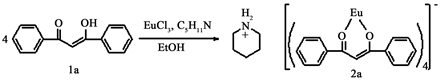
Scheme 1. Preparation of piperidinium tetrakis(1,3-diphenyl-1,3-propanedionato) europate (III).

Figure 1. UV-Vis spectra of complex 2a.

Figure 2. Emission spectra of the complex 2a.
with 5D0 → 7FJ (J = 0 - 4) transitions of the europium (III) ion. The very high intensity peak was observed at 613 nm due to the 5D0 → 7F2 transition, suggesting a highly polarizable chemical environment around the europium (III) ion [18] . This transition was responsible for the red emission of the complex 2a.
Suitable single crystals for X-ray structure analysis were easily obtained for the complex 2a. In principle, europium (III) complexes are not sensitive against air, therefore, preparation and purification of the complex is quite easy. Crystallization of the complex 2a was carried out using three different solvents (chloroform, ethanol, and benzene) that resulted in crystals of complex 2a and its other two crystals 2b, 2c having solvent molecules in their cavity.
Molecular shapes of these complexes are fused dual-cone like structures and the complex molecules have cavities (Figure 3). The complex 2a crystallizes in the monoclinic space groups Cc (centrosymmetric, No.13), the cell unit includes four molecules with four piperidinium ions and with no solvate molecule (Figure 3).


Figure 3. ORTEP view of the complexes 2a (left), 2b (center) and 2c (right). Ellipsoids are drawn at 50% probability level. Aqua blue, blue and red ellipsoids show Eu, N and O atom(s), respectively.
The complex 2b crystallizes in the monoclinic space groups P21/n (centrosymmetric, No.14); the cell unit includes four molecules with four piperidinium ions and with eight water molecules (Figure 3). The complex 2c also crystallizes in the monoclinic space group P21/n (centrosymmetric, No.14), the cell unit includes four molecules with four piperidinium ions and with four benzene molecule (Figure 3). The three crystals have quite complex molecular packing arrangement (Figure 4).
The europium (III) ions of 2a, 2b and 2c are coordinated by a distorted octahedral arrangement of eight oxygen atoms from four chelating β-diketonato ligands. The average Eu1-O (1 - 8) bond lengths are moderately normal, and these values are ca.2.378 Å for 2a, 2.389 Å for 2b and 2.391 Å for 2c, respectively. The bond angles in the five membered rings consisting of Eu and 1,3-butanedionato ligands (O-Eu-O) vary from 69.7 (2)˚ to 71.7 (1)˚ for 2a, 68.9 (3)˚ to 71.3 (3)˚ for 2b and 70.45 (5)˚ to 71.01 (5)˚ for 2c, respectively (Tables 2-4). These values of bond distances and bond angles are in good agreement with those reported for other analogous Eu-β-diketonato complexes [19] . The piperidinium cation involving the N1atomis most stable chair form in the crystal.
Molecular shapes of these complexes are fused double-cone like structures and the complex molecules have cavities (Figure 3). The difference between 2a, 2b and 2c about the inclusion of solvate molecule is well explained that the difference of the largeness of the cavity consisting of the four ligands. The cavity


Figure 4. Crystal packing diagram of the complexes 2a (above) and 2b (below). Aqua blue, pale green, darkblue, red and green ellipsoids show Eu, F, N, O and Cl atoms, respectively.
Table 2. Selected bond lengths (Å) and angles (˚) for the complex 2a.
Table 3. Selected bond lengths (Å) and angles (˚) for the complex 2b.
Table 4. Selected bond lengths (Å) and angles (˚) for the complex 2c.
sizes of the complex 2a seems to be larger. The large cavity sizes of the complex 2a facilitated the inclusion of water and benzene solvate molecules. Differences between these complexes and Calix [4] arene are environments internal spaces of cavities, in another words, aromatic parts on the Calixarenes are oriented π-electron surfaces to the cavity center. On the contrary, the aromatic parts on the complexes are oriented aromatic C-H protons to the cavity center. Therefore, the complex 2a, expected as new compound for undeveloped field of host-guest chemistry.
4. Conclusion
In conclusion, we reported the synthesis, characterization, crystal structure and photophysical properties of piperidinium [tetrakis(1,3-diphenyl-1,3-propanedionato) europate (III)] complex 2a. Its two different crystals 2b, 2c having water, benzene solvent molecules were readily prepared by crystallization process. Complex 2a was characterized by 1H-NMR spectroscopies, positive FAB-Mass, and Elemental Analysis. Further their exact molecular structures of 2a, 2b and 2c were determined by X-ray analysis. The UV-Vis, Fluorescence properties of all three crystals remained same in solid as well as in solution state. Complex 2a exhibited strong red emission at 613 nm, which could find prominent applications in light emitting devices. These strong emissions were attributed to the 5D0 → 7F2 transition of Europium (III) ions under UV excitation.
Acknowledgements
We are grateful to the Center for Instrumental Analysis, Kyushu Institute of Technology (KITCIA) for the electron impact mass, 1HNMR spectra and X-ray analysis. This research was financially supported by JSPS KAKENH Grant Number 15K05611
Cite this paper
Moriguchi, T., Kitou, N., Yakeya, D., Tsuge, A., Yoza, K. and Jalli, V. (2017) Syntheses, Crystal Structure and Photoluminescence Properties of Piperidinium Tetrakis (1,3-Diphenyl-1,3-Propanedionato) Europate(III) Complex and Its Two Different Crystals. Crystal Structure Theory and Applications, 6, 57-66. https://doi.org/10.4236/csta.2017.64005
References
- 1. Pietraszkiewicz, O., Mal, S., Pietraszkiewicz, M., Maciejczyk, M., Czerski, I., Borowiak, T., Dutkiewicz, G., Drobchak, O., Penninck, L., Beeckman, J. and Neyts, K. (2012) Highly Photoluminescent Eu (III) Complexes of the New 1-Triphenylen-2-yl-3- Trifluoroacetylacetone. Journal of Photochemistry and Photobiology A, 250, 85-91. https://doi.org/10.1016/j.jphotochem.2012.10.003
- 2. De Sá, G.F., Malta, O.L., De Mello Donegá, C., Simas, A.M., Longo, R.L., Santa-Cruz, P.A. and Da Silva, E.F. (2000) Spectroscopic Properties and Design of Highly Luminescent Lanthanide Coordination Complexes. Coordination Chemistry Reviews, 196, 165-195. https://doi.org/10.1016/S0010-8545(99)00054-5
- 3. Zhang, Y., Shi, H., Ke, Y. and Cao, Y. (2007) Synthesis and Characterization of Highly Fluorescent Europium Functionalized β-Diketonate Complexes. Journal of Luminescence, 124, 51-57. https://doi.org/10.1016/j.jlumin.2006.01.361
- 4. Hasegawa, Y., Wada, Y. and Yanagida, S. (2004) Strategies for the Design of Luminescent Lanthanide (III) Complexes and Their Photonic Applications. Journal of Photochemistry and Photobiology C, 5, 183-202. https://doi.org/10.1016/j.jphotochemrev.2004.10.003
- 5. Parker, D., Senanayake, P.K. and Gareth Williams, J.A. (1998) Luminescent Sensors for pH, pO2, Halide Hydroxide Ions Using Phenanthridine as a Photosensitiser in Macrocyclic Europium and Terbium Complexes. Journal of the Chemical Society, Perkin Transactions 2, 10, 2129-2139. https://doi.org/10.1039/a801270i
- 6. Robinson, M.R., O’Reganc, M.B. and Bazan, G.C. (2000) Synthesis, Morphology and Optoelec Tronic Properties of Tris [(N-Ethylcarbazolyl)(3A, 5A Hexyloxybenzoyl) me Thane](Phenanthroline) Europium. Chemical Communications, 1645-1646. https://doi.org/10.1039/b004739m
- 7. Aromi, G., Gamez, P. and Reedijk, J. (2008) Poly Beta-Diketones: Prime Ligands to Generate su Pramolecular Metalloclusters. Coordination Chemistry Reviews, 252, 964-989.
- 8. Manju, B., Satish, K., Taxak, V.B., Priti, B. and Khatkar, S.P. (2015) Synthesis, Photolumines Cent Features and Intramolecular Energy Transfer Mechanism of Europium (III) Complexes with Fluorinate b-Diketone Ligand and Auxiliary Ligands. Journal of Fluorine Chemistry, 178, 6-13.
- 9. Wang, D., Liu, H., Fan, L., Yin, G., Hu, Y. and Zheng, J. (2015) Synthesis and Photolumines Cent Behavior of Eu(III) Complexes with 4,4,4-trifluoro-1-(6-methoxy naphthalen-2-yl)-butane-1,3-dione. Synthetic Metals, 209, 267-272.
- 10. Kalinovskaya, I.V. and Mirochnik, A.G. (2015) Luminescent Properties of Compounds of Europium(III) with Quinaldic Acid and β-Diketones. Optics and Spectroscopy, 119, 992-995. https://doi.org/10.1134/S0030400X15110119 https://link.springer.com/article/10.1134/S0030400X15110119
- 11. Malba, C.M., Enrichi, F., Facchin, M., Demitri, N., Plaisier, J.R., Natile, M.M., Selva, M., Ri ello, P., Perosa, A. and Benedetti, A. (2015) Phosphonium-Based Tetrakis Dibenzoylmethane Eu(III) and Sm(III) Complexes: Synthesis, Crystal Structure and Photoluminescence Properties in a Weakly Coordinating Phosphonium Ionic Liquid. RSC Advances, 5, 60898-60907. https://doi.org/10.1039/C5RA03947A
- 12. Martins, J.P., Martín-Ramos, P., Coya, C., Silva, M.R., Eusebio, M.E.S., deAndrés, A., álva rez, A.D. and Martín-Gil, J. (2015) Highly Luminescent Pure-Red-Emitting Fluorinated β-Diketonate Europium(III) Complex for Full Solution-Processed OLEDs. Journal of Luminescence, 159, 17-25.
- 13. Moriguchi, T., Hirosaki, S., Venkataprasad, J., Tsuge, A. and Yoza, K. (2017) The Syntheses, Crystal Structure and Luminescence Properties of Cone-Like Octadentate Europium (III) Complexes with Four Short Alkoxy Substituents. Crystals, 7, 85-93. https://doi.org/10.3390/cryst7030085
- 14. Bruker AXS Inc. (2009) APEX2 Version 2009.9. Tokyo.
- 15. Bruker AXS Inc. (2009) SAINT Version 7.68A. Tokyo.
- 16. Sheldrick, G.M.A. (2008) Short History of SHELX. Acta Cryst A, 64, 112-122. https://doi.org/10.1107/S0108767307043930
- 17. Melby, L.R., Rose, N.J., Abramson, E. and Caris, J.C. (1964) Synthesis and Fluorescence of Some Trivalent Lanthanide Complexes. Journal of the American Chemical Society, 86, 5117-5125. https://doi.org/10.1021/ja01077a015
- 18. Richardson, F.S. (1982) Terbium(III) and Europium(III) Ions as Luminescent Probes and Stains for Biomolecular Systems. Chemical Reviews, 82, 541-552. https://doi.org/10.1021/cr00051a004
- 19. Zhang, L., Li, B., Zhang, L., Chen, P. and Liu, S. (2009) Synthesis, Characterization, and Lu Minescent Properties of Europium Complexes with Fluorine Functionalized Phenanthroline. Journal of The Electrochemical Society, 156, H202-H207. https://doi.org/10.1149/1.3060228