Open Journal of Veterinary Medicine
Vol.3 No.4(2013), Article ID:35739,5 pages DOI:10.4236/ojvm.2013.34037
Detection of Antibodies in Serum Specimens from Dogs with Blastomycosis with Lysate Antigens Prepared from Four Blastomyces dermatitidis Dog Isolates: Individual Antigens vs Antigen Combinations
Department of Biological Sciences, Idaho State University, Pocatello, USA
Email: vandjami@isu.edu
Copyright © 2013 Jamie L. VanDyke et al. This is an open access article distributed under the Creative Commons Attribution License, which permits unrestricted use, distribution, and reproduction in any medium, provided the original work is properly cited.
Received June 13, 2013; revised July 13, 2013; accepted July 26, 2013
Keywords: Blastomycosis; Lysate Antigen Combinations; Antibody Detection; ELISA
ABSTRACT
Blastomycosis, the systemic fungal infection of humans and animals, has presented a diagnostic challenge to clinicians and laboratory personnel for many years. Our laboratory has been concentrating on attempting to develop antigenic reagents from the yeast phase of various isolates of Blastomyces dermatitidis and to evaluate these lysate antigens with regard to antibody detection in blastomycosis. The aim of this current study was to evaluate yeast phase antigens prepared from four dog isolates of B. dermatitidis and to evaluate their efficacy, when used individually or in combination, for antibody detection in sera from dogs with blastomycosis. Mean absorbance values using the ELISA to assay 24 serum specimens (Trial 1) ranged from 0.588 with an individual lysate antigen to 0.992 when three reagents were combined. Eight of the lysates exhibited mean absorbance values ranging from 0.992 to 0.915 with 7 out of 8 being lysate antigen combinations. Mean absorbance values with the other 6 lysates ranged from 0.899 to 0.588. In Trial 2, the 6 most sensitive reagents from Trial 1 were assayed against 10 highly reactive dog sera. The results of Trial 2 showed that 5 antigen combinations detected antibody to a greater degree than the individual lysate antigen. Combinations of northern and southern antigens were able to detect antibody in serum specimens from either of these geographical regions. Comparative studies are continuing to further evaluate various lysate antigen combinations for antibody detection in blastomycosis.
1. Introduction
The systemic fungal disease, blastomycosis, caused by Blastomyces dermatitidis, occurs in humans and other animals. The disease is found mainly in the United States in southeastern, south-central and upper Midwestern states and the fungus exists as a soil saprophyte associated with slightly acidic soils and often found near a water source [1-3].
The infection is initiated by the inhalation of the mycelial spore. The thermally dimorphic organism exists in this stage in nature or in the laboratory at 25˚C and has the ability to convert to the yeast phase at 37˚C in the lungs of the infected host and produces a primary pulmonary infection. Blastomycosis may be self-resolving or produce an acute or chronic disease state in the lungs. In some cases the organism may disseminate to other organs including the central nervous system with the possible development of fatal meningitis [4-10].
The diagnosis of the disease has presented major problems to clinicians and laboratory personnel for many years. Culturing of the organism or histopathologic methods have been used, but these procedures may be time consuming. They also may not produce a reliable diagnosis or in some cases a misdiagnosis [2,5,7-9]. The use of immunodiagnostic assays has been very successful with many infectious diseases, but assays that have been developed for antibody detection in blastomycosis have been hampered by problems with sensitivity and specificity [5,7-11]. In recent years investigators have continued to put a considerable amount of efforts into developing reliable immunodiagnostic assays for the disease and have contributed with novel antibody and antigen detection systems [8,11-17].
For the past several years our laboratory has been involved in the production of B. dermatitidis yeast-phase lysate antigens from human, animal and environmental isolates of the fungus and in performing various comparative studies on the lysates with regard to antibody detection in serum specimens from immunized and infected animals or the use of these lysate antigens as immunizing agents and the subsequent use of the antibodies for antigen detection assays. Encouraging results have been obtained by using various enzyme immunoassays (ELISAs) to compare the lysate reagents in antibody or antigen detection assays [18-28].
Our research continues to emphasize that we need to learn more about strain differences of the fungus and how these differences may influence the development of improved methods for the immunodiagnosis of blastomycosis in humans and animals. We have performed numerous comparative studies on individual lysate reagents, but little with respect to evaluating antigen combinations for antibody detection. The current study was designed to compare yeast lysate antigens prepared from 4 B. dermatitidis dog isolates (Tennessee and Wisconsin) for antibody detection in serum specimens from dogs with diagnosed blastomycosis. The 4 lysates were tested individually and in combinations (14 antigenic preparations) to determine sensitivity parameters.
2. Materials and Methods
2.1. Lysate Antigen Preparation
Yeast phase lysate reagents (T-58, dog Tennessee, T-66, dog Tennessee, WI-R, dog Wisconsin and WI-J, dog Wisconsin) were prepared by a method similar to one that was previously used for the production of antigen from Histoplasma capsulatum [29,30] and modified in our laboratory for B. dermatitidis lysate antigen production [18]. The yeast phase cells were grown for 7 days at 37˚C in a chemically defined medium in an incubator shaker, harvested by centrifugation (700 × g; 5 min), followed by washing with distilled water, resuspended in distilled water and then allowed to lyse for 7 days at 37 C in water with shaking. The preparations were centrifuged, filter sterilized, merthiolate added (1:10,000) and stored at 4˚C. Protein determinations were performed on the lysates using the BCA Protein Assay Kit (Pierce Chemical Company, Rockford, IL) and dilutions of the antigenic reagents used in the ELISAs assays were based on protein concentration. Combinations of the above four antigenic reagents were also used to detect antibodies, as indicated below.
2.2. Serum Specimens
Twenty-four serum specimens from dogs with diagnosed blastomycosis from both northern and southern regions of the United States were provided by Dr. A.M. Legendre (University of Tennessee College of Veterinary Medicine, Knoxville, TN).
2.3. Enzyme-linked Immunosorbent Assay (ELISA)
The ability of each of the 14 (individual or combination preparations) yeast lysate reagents to detect antibodies in the above serum specimens was determined using the indirect enzyme-linked immunosorbent assay (ELISA). Each lysate antigen was diluted (2000 ng/ml of protein) in a carbonate-bicarbonate coating buffer (pH 9.6) and then added to triplicate wells (100 ul) of a NUNC 96-well microplate (Fisher-Thermo). The plates were then incubated overnight at 4˚C in a humid chamber followed by washing three times with phosphate buffered saline containing 0.15% Tween 20 (PBS-T). The serum specimens (1:2500 dilution; 100 ul) were added to the microplate wells and incubated for 30 min at 37˚C in a humid chamber. Following this incubation the wells were washed as above and 100 ul of goat anti-dog IgG (H & L) peroxidase conjugate (Kirkegaard and Perry, Gaithersburg, MD) was added to each well and incubated for 30 min at 37˚C. The plates were again washed as above and 100 ul of TMB peroxidase substrate (Pierce/Fisher-Thermo) was added to each well and incubated for approximately 2 min at room temperature. The reaction was stopped by the addition of sulfuric acid and the absorbance read at 450 nm using a BIO-RAD 2550 EIA reader.
3. Results
The mean absorbance values of the 14 B. dermatitidis lysate antigens, when used in the ELISA to detect antibodies in 24 dog sera, are shown in Table 1. Six of the reagents exhibited absorbance values ranging from 0.933 to 0.992 with a mean value of 0.961. Seven of the 8 reactive preparations were antigen combinations; the most reactive reagent was composed of antigens T-66 (a Tennessee isolate) and WI-J and WI-R (Wisconsin isolates). When the mean absorbance values and the median absorbance values of the individual antigens and antigen combinations obtained in Trial 1 were compared, it showed that 6 of the antigenic reagents were optimal, (based on mean or median determinations). Therefore it was decided to further evaluate the 6 preparations against 10 highly reactive sera in order to determine if variations in antibody content in the 24 serum specimens might possibly influence overall mean/median absorbance values of the antigens.
Figures 1 and 2 represent the efficacy of the 14 antigenic preparations in Table 1 when used for antibody detection in the serum specimens from the 24 dogs with blastomycosis (Mean abs. value range of 0.588 to 0.992).
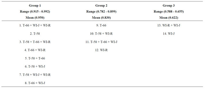
Table 1. Mean absorbance values obtained when the 14 Blastomyces dermatitidis yeast lysate antigens/antigen combinations were used to detect antibodies in 24 dog serum specimens from northern (Minnesota) and southern (Tennessee, Alabama, Louisiana) regions of the U.S.
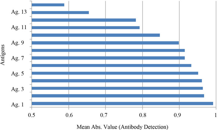
Figure 1. Comparison of the reactivity (Mean Abs. Values) of the 14 B. dermatitidis yeast lysate antigens with respect to antibody detection in 24 dog serum specimens (Trial 1). The antigens (1 - 14) refer to the lysate preparations shown above in Table 1.
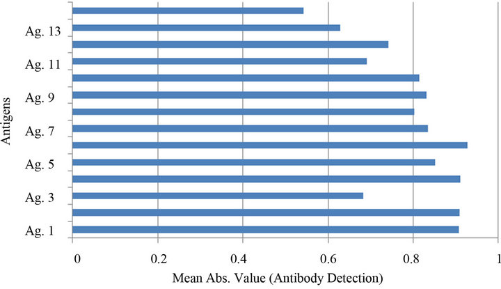
Figure 2. Comparison of the reactivity (Median Abs. Values) of the 14 B. dermatitidis yeast lysate antigens with respect to antibody detection in 24 dog serum specimens (Trial 1). The antigens (1 - 14) refer to the lysate preparations shown above in Table 1.
Several of the reagents were able to detect antibodies in the sera in a sensitive manner, but the 6 with the greatest degree of reactivity were (1) T-66 + WI-J + WI-R, (2) T-58, (3) T-58 + T-66 + WI-R, (4) T-66 + WI-R , (5) T-58 + T-66 and (6) T-58 + WI-J (mean absorbance value of the above six most reactive antigens was 0.961).
The results of Trial 2 (Figure 3), when the 6 lysate preparations which exhibited the greatest reactivity in Trial 1were assayed against 10 highly reactive dog sera, indicated that antigen 3 (T-58 + T-66 + WI-R) was optimal with a mean absorbance value of 1.387. The reactivity of the other antigens ranged from 1.364 (antigen 4; T-66 + WI-R) to 1.184 (antigen 2; T-58). In this evaluation the greatest reactivity was obtained with the 5 north/ south antigen combinations while the individual antigen preparation (T-58) was slightly less reactive.
Based on these results, it appears that the 6 most reactive antigenic reagents might be divided into 3 groups: A: highly reactive (T-58 + T-66 + WI-R and T-66 + WI-R), B: moderately reactive (T-58 + WI-J; T-66 + WI-J + WI-R and T-58 + T-66) and C: less reactive (T-58). Therefore, any of the six preparations could reliably detect antibodies in dogs with blastomycosis, but the two highly reactive reagents would be the antigens of choice for antibody detection in this study.
4. Discussion/Conclusions
When developing antigenic reagents for the immunodiagnosis of blastomycosis or other infectious diseases, it is desirable that the diagnostic antigens are able to detect antibody in humans and animals from a variety of geo-
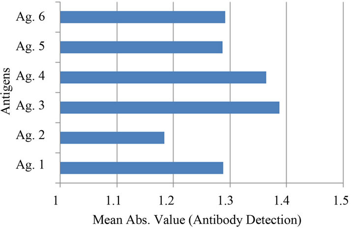
Figure 3. Comparison of the 6 B. dermatitidis yeast lysate antigens exhibiting the greatest reactivity in Trial 1 when assayed against 10 dog serum specimens. The antigens (1 - 6) refer to the lysate preparations shown above in Table 1.
graphical regions of the U.S or their countries. Our laboratory has been concerned for the past several years with developing and evaluating novel B. dermatitidis yeast phase lysate antigens prepared from isolates of the fungus from human, animal and environmental sources and the use of such antigens for antibody detection or producing antibodies from the antigens for antigen detection [18-28].
Most of these studies have been involved with the use of lysate antigens prepared from one isolate of the organism, but we have recently become interested in evaluating combinations of antigens prepared from different isolates of B. dermatitidis in order to achieve greater antibody detection. The present study was designed to evaluate lysates prepared from 4 different isolates (individually and in combinations) to detect antibodies in sera from dogs with blastomycosis. All of the 14 antigenic reagents were able to detect antibody (mean absorbance range of 0.588 to 0.992) in the dog sera, but the most reactive reagents were combinations of the T-58, T-66, WI-J and WI-R lysate antigens.
The results of Trial 1 and Trial 2 seem to indicate that the combination of antigens prepared from B. dermatitidis isolates from both the north (Wisconsin) and south (Tennessee) are efficient as immunodiagnostic reagents for antibody detection in sera from dogs from both northern (Minnesota) and southern (Tennessee, Alabama, Louisiana) regions of the U.S. The data provided by this study is certainly encouraging with regard to further evaluations of combinations of lysate antigens prepared from isolates of B. dermatitidis from various sources. Therefore, we are continuing such studies in an effort to develop an optimal antigen(s) for the reliable diagnosis of blastomycosis in animals and humans.
5. Acknowledgements
This research was supported by the Department of Biological Sciences, Idaho State University, Pocatello, ID 83209-8007.
REFERENCES
- M. A. Pfaller and D. J. Diekema, “Epidemiology of Invasive Mycoses in North America,” Critical Reviews in Microbiology, Vol. 36, No.1, 2010, pp. 1-53. doi:10.3109/10408410903241444
- A. F. DiSalvo, “Blastomycosis, in Topley and Wilson’s Microbiology and Microbial Infections,” 9th Edition, Arnold Publishers, London, 1998.
- M. A. DeGroote, R. Bjerke, H. Smith and L. V. Rhodes III, “Expanding Epidemiology of Blastomycosis: Clinical Features and Investigation of 2 Cases in Colorado,” Clinical Infectious Diseases, Vol. 30, No. 3, 2000, pp. 582- 584. doi:10.1086/313717
- R. W. Bradsher, “Clinical Features of Blastomycosis,” Seminars in Respiratory Infections, Vol. 12, No. 3, 1997, pp. 229-234.
- R. W. Bradsher, S. W. Chapman and P. G. Pappas, “Blastomycosis,” Infectious Disease Clinics of North America, Vol.17, No. 1, 2003, pp. 21-40. doi:10.1016/S0891-5520(02)00038-7
- J. A. Smith and C. A. Kauffman, “Pulmonary Fungal Infections,” Respirology, Vol. 17, No. 6, 2012, pp. 913- 926. doi:10.1111/j.1440-1843.2012.02150.x
- J. R. Bariola and K. S. Vyas, “Pulmonary Blastomycosis,” Seminars in Respiratory Critical Care Medicine, Vol. 32, No. 6, 2011, pp. 745-753. doi:10.1055/s-0031-1295722
- J. A. McKinnell and P. G. Pappas, “Blastomycosis: New Insights into Diagnosis, Prevention, and Treatment,” Clinical Chest Medicine, Vol. 30, No. 2, 2009, pp. 227- 239. doi:10.1016/j.ccm.2009.02.003
- M. Saccente and G. L. Woods, “Clinical and Laboratory Update on Blastomycosis,” Clinical Microbiology Reviews, Vol. 23, No. 2, 2010, pp. 367-381. doi:10.1128/CMR.00056-09
- J. E. Cutler, G. S. Deepe Jr. and B. S. Klein, “Advances in Combating Fungal Diseases: Vaccines on the Threshold,” Nature Review of Microbiology, Vol. 5, No. 1, 2007, pp. 13-18. doi:10.1038/nrmicro1537
- K. S. Vyas, J. R. Bariola and R. W. Bradsher, “Advances in the Serodiagnosis of Blastomycosis,” Current Fungal Infection Reports, Vol. 2, No. 4, 2008, pp. 227-231. doi:10.1007/s12281-008-0033-z
- B. S. Klein and J. M. Jones, “Isolation, Purification and Radiolabeling of a Novel Surface Protein on Blastomyces dermatitidis Yeasts to Detect Antibody in Infected Patients,” Journal of Clinical Investigation, Vol. 85, No. 1, 1990, pp. 152-161. doi:10.1172/JCI114406
- P. Connolly, C. A. Hage, J. R. Bariola, E. Bensadoun, M. Rodgers, R. W. Bradsher and J. J. Wheat, “Blastomyces dermatitidis Antigen Detection by Quantitative Enzyme Immunoassay,” Clinical Vaccine Immunology, Vol. 19, No. 1, 2012, pp. 53-56. doi:10.1128/CVI.05248-11
- C. A. Hage, T. E. Davis, L. Egan, M. Parker, D. Fuller, A. M. LeMonte, D. Durkin, P. Connelly, L. J. Wheat, D. BlueHindy and K. A. Knox, “Diagnosis of Pulmonary Histoplasmosis and Blastomycosis by Detection of Antigen in Bronchoalveolar Lavage Fluid Using an Improved Second-Generation Enzyme-Linked Immunoassay,” Respiratory Medicine, Vol. 101, 2007, pp. 43-47. doi:10.1016/j.rmed.2006.04.017
- B. S. Klein, R. A. Squires, J. K. Lloyd, D. R. Ruge and A. M. Legendre, “Canine Antibody Response to Blastomyces dermatitidis WI-1 Antigen,” American Review of Veterinary Research, Vol. 61, No. 5, 2000, pp. 554-558. doi:10.2460/ajvr.2000.61.554
- D. Spector, A. M. Legendre, J. Wheat, D. Bemis, B. Rohrbach, J. Taboada and M. Durkin, “Antigen and Antibody Testing for the Diagnosis of Blastomycosis in Dogs,” Journal of Veterinary Internal Medicine, Vol. 22, No. 4, 2008, pp. 839-843. doi:10.1111/j.1939-1676.2008.0107.x
- J. R. Bariola, C. A. Hage, M. Durkin, E. Bensadoun, P. O. Gubbins, L. J. Wheat and R. W. Bradsher, “Detection of Blastomyces dermatitidis Antigen in Patients with Newly Diagnosed Blastomycosis,” Diagnostic Microbiology and Infectious Diseases, Vol. 69, No. 2, 2011, pp. 187-191. doi:10.1016/j.diagmicrobio.2010.09.015
- S. M. Johnson and G. M. Scalarone, “Preparation and ELISA Evaluation of Blastomyces dermatitidis Yeast Phase Lysate Antigens,” Diagostic Microbiology and Infectious Diseases, Vol. 11, No. 2, 1989, pp. 81-86. doi:10.1016/0732-8893(88)90076-4
- J. L. Bono, A. M. Legendre and G. M. Scalarone, “Detection of Antibodies and Delayed Hypersensitivity with Rotofor Preparative IEF Fractions of Blastomyces dermatitidis Yeast Phase Lysate Antigens,” Journal of Medical and Veterinary Mycology, Vol. 33, No. 4, 1995, pp. 209- 214. doi:10.1080/02681219580000441
- M. A. Fisher, J. L. Bono, R. O. Abuodeh, A. M. Legendre and G. M. Scalarone, “Sensitivity and Specificity of an Isoelectric Focusing Fraction of Blastomyces dermatitidis Yeast Lysate Antigen for the Detection of Canine Blastomycosis,” Mycoses, Vol. 38, No. 5-6, 1995, pp. 177- 182. doi:10.1111/j.1439-0507.1995.tb00046.x
- R. C. Axtell and G. M. Scalarone, “Serological Differences in Three Blastomyces dermatitidis Strains,” Mycoses, Vol. 45, No. 11-12, 2002, pp. 437-422. doi:10.1046/j.1439-0507.2002.00804.x
- J. F. Shurley, A. M. Legendre and G. M. Scalarone, “Blastomyes dermatitidis Antigen Detectioni in Urine Specimens from Dogs with Blastomycosis Using a Competitive Binding Inhibition ELISA,” Mycopathologia, Vol. 160, No. 2, 2005, pp. 137-142. doi:10.1007/s11046-005-3153-9
- J. F. Shurley and G. M. Scalarone, “Isoelectric Focusing and ELISA Evaluation of a Blastomyces dermatitidis Human Isolate,” Mycopathologia, Vol. 164, No. 2, 2007, pp. 73-76. doi:10.1007/s11046-007-9033-8
- C. M. Sestero and G. M. Scalarone, “Detection of IgG and IgM in Sera from Canines with Blastomycosis Using Eight Blastomyces dermatitidis Yeast Phase Lysate Antigens,” Mycopathologia, Vol. 162, No. 1, 2006, pp. 33-37. doi:10.1007/s11046-006-0028-7
- C. M. Sestero and G. M. Scalarone, “Detection of the Surface Antigens BAD-1 and Alpha (1-3) Glucan in Six Different Strains of Blastomyces dermatitidis Using Monoclonal Antibodies,” Journal of Medical and Biological Science, Vol. 1, No. 1, 2006, pp. 1-7.
- W. O. Hatch and G. M. Scalarone, “Comparison of Colorimetric and Chemiluminescent ELISAs for the Detection of Antibodies to Blastomyces dermatitidis,” Journal of Medical and Biological Sciences, Vol. 3, No 1, 2009, pp. 1-6.
- J. C. Wright, T. E. Harrild and G. M. Scalarone, “Sensitivity and Specificity Determinations with Isoelectric Focusing Fractions of Blastomyces dermatitidis for Antibody Detection in Serum Specimens from Infected Dogs,” Open Journal of Vetrinary Meddicine, Vol. 2, No. 4, 2012, pp. 237-241. doi:10.4236/ojvm.2012.24038
- J. J. Roberts, M. V. Madrid, L. Dickerson, B. Hutchison and G. M. Scalarone, “Comparison of Antibody Detection with Yeast Lysate Antigens Prepared from Blastomyces dermatitidis Dog Isolates from Wisconsin and Tennessee,” Open Journal of Veterinary Medicine, Vol. 3, No. 1, 2013, pp. 67-72. doi:10.4236/ojvm.2013.31011
- H. B. Levine, G. M. Scalarone and S. D. Chaparas, “Preparation of Fungal Antigens and Vaccines: Studies on Coccidioides immitis and Histoplasma capsulatum,” Contributions to Microbiology and Immunology, Vol. 3, 1977, pp. 106-125.
- G. M. Scalarone, H. B. Levine and S. D. Chaparas, “Delayed Hypersensitivity Responses of Experimental Animals to Histoplasmin from the Yeast and Mycelial Phases of Histoplasma capsulatum,” Infection and Immunity, Vol. 21, No. 3, 1978, pp. 705-713.