Open Journal of Gastroenterology
Vol.2 No.2(2012), Article ID:19132,2 pages DOI:10.4236/ojgas.2012.22007
Epiploic appendagitis as an uncommon cause of lower abdominal pain
![]()
1Department of General Medicine, Asahikawa Medical University, Asahikawa, Japan
2Department of Medicine, Asahikawa Medical University, Asahikawa, Japan
3Department of Regional Medicine and Education, Asahikawa Medical University, Asahikawa, Japan
Email: *okumurat@asahikawa-med.ac.jp
Received 22 March 2012; revised 12 April 2012; accepted 25 April 2012
Keywords: Epiploic Appendagitis; Lower Abdominal Pain; CT
ABSTRACT
Epiploic appendagitis should be considered to be an uncommon cause of lower abdominal pain. To diagnose accurately, typical CT findings are needed, and total colonoscopy should be done later to rule out the possibility of diverticulosis.
1. CASE REPORT
A 33-year-old Japanese man presented to the outpatient department with acute left lower abdominal pain. He had no fever and no other associated symptoms. On examination, he was tender in the left lower abdominal quadrant. Routine blood investigations were normal except mild elevation of C-reactive protein (1.12 mg/ml). The presumptive diagnosis after history taking and physical examination was diverticulitis. The patient underwent computer tomography of the abdomen, which showed an oval lesion, maximum diameter 3.0 cm, with fat attenuation, located adjacent to the descending colon (Figure 1(b), circle), and the diagnosis of primary epiploic appendagitis (PEA) was strongly indicated. To exclude the possibility that the cause was diverticulitis, colonoscopy was performed a couple of weeks later, and demonstrated that there was no diverticulosis. PEA is a rare condition that results from inflammation of an epiploic appendage by spontaneous torsion or a hemorrhagic infarct and is an often misdiagnosed cause of acute abdominal pain, making it an important differential diagnosis [1]. The epiploic appendages are peritoneal pouches that arise from the serosal surface of the colon originating next to the anterior and the posterior taenia coli. Usually their sizes are 1 - 2 cm in thickness and 0.5 - 5 cm in length. Approximately 50 - 100 epiploic appendages are distributed from the cecum to the rectosigmoid junction. Depending on its location, it can mimic many disorders such as colonic diverticulitis, acute appendicitis, a gynaecological disorder or acute cholecystitis. CT has been reported to be a reliable detection for PEA [1]. Since the treatment for PEA should be conservatively with or without anti-inflammatory drugs usually sufficient to control pain and no surgical intervention is needed because PEA is spontaneously resolved [1]. The present patient was managed conservatively and recovered well within a week. Since he had visited to our clinic 6 months before to examine the cause of hematuria, CT scan at the abdomen was then performed. Figure 1 also illustrates CT imagines of the abdomen approximately 6 months before (Figure 1(a)) and 2 months after (Figure 1(c)) the diagnosis of epiploic appendagitis for follow-up. As clearly demonstrated, the oval lesion was hardly detected at the estimated location at both time points, suggesting that the 3 cm-oval lesion appeared and then disappeared within a couple of months. In normal conditions, epiploic appendages are not detectable on a CT scan. It has been also described that after an appendage becomes necrotic, the nonviable appendage is absorbed by the body. It has also been suggested that detachment of epiploic appendages might be a source of loose intraperitoneal bodies,
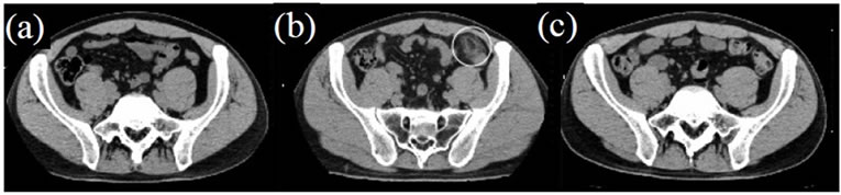
Figure 1. Computer tomography (CT) of the abdomen showed an oval lesion, maximum diameter 3.0 cm, with fat attenuation, located adjacent to the descending colon, strongly indicating primary epiploic appendagitis (b, circle), but the oval lesion was hardly detected at the estimated location at both time points such as approximately 6 months before (a) and 2 months after (c).
which are found incidentally by laparoscopy [2,3]. These evidence are in good agreement with the time-course change of the CT findings of PEA as seen in this case. In the past, the standard treatment for PEA was surgical excision because it was diagnosed during laparotomy in most cases [4,5]. However, as described in recent papers, PEA could be treated without surgical operation [1]. We therefore can not confirm the lesion is indeed PEA pathologically. Considering the clinical characteristics of PEA as seen in this case, we need to show to get an accurate diagnosis that typical CT findings of epiploic appendagitis would disappear during the follow-up period, and no diverticulosis of the colon should be proved by colonoscopy.
REFERENCES
- Schnedl, W.J., Krause, R., Tafeit, E., et al. (2011) Insights into epiploic appendagitis. Nature Reviews Gastroenterology & Hepatology, 8, 45-49.
- Borg, S.A., Whitehouse, G.H. and Griffiths, G.J. (1976) A mobile calcified amputated appendix epiploica. American Journal of Roentgenology, 127, 349-350.
- Elliot, G.B. and Freigang, B. (1962) Aspetic necrosis, calcification and separation of appendices epiploicae. Annals of Surgery, 155, 501-505.
- Shehan, J.J., Organ, C. and Sullivan, J.F. (1966) Infarction of the appendices epiploicae. The American Journal of Gastroenterology, 46, 469-476.
- Carmichael, D.H. and Organ, C.H. (1985) Epiploic disorders: Conditions of the epiploic appendages. Archives of Surgery, 120, 1167-1172. doi:10.1001/archsurg.1985.01390340063012
NOTES
*Corresponding author.